
 Goat Anti-Rabbit IgG (H&L) - AcalephFluor790
  Datasheet
 MSDS
MSDS
Description: Goat Polyclonal Secondary Antibody to Rabbit IgG (H&L) - AcalephFluor790 Immunogen: Rabbit IgG Purification: The antibody was isolated from antisera by immunoaffinity chromatography using antigens coupled to agarose beads. Clonality: Polyclonal Conjugation: AcalephFluor790 Form: Liquid in 0.01M Phosphate Buffered Saline, pH 7.2, containing 1% BSA, 50% glycerol, 0.02% Sodium Azide Dilution: WB (1/5000 - 1/20000), IH (1/100 - 1/1000), IF (1/100 - 1/1000), FC (1/1000 - 1/4000), E (Use at an assay dependent concentration) Storage/Stability : Shipped at 4°C. Upon delivery aliquot and store at -20°C for one year. Avoid freeze/thaw cycles.
Platform : Ex/Em = 770/794 nm
-
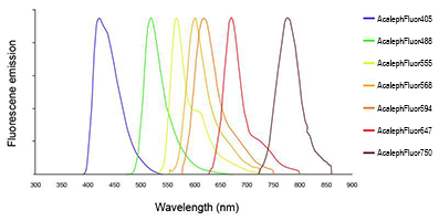 Line colors represent the approximate visible colors of the wavelength maxima.
 -
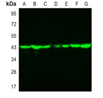 Western blot analysis of Beta-actin expression in Jurkat (A), MCF7 (B), NIH3T3 (C), mouse brain (D), mouse liver (E), rat brain (F), rat liver (G) whole cell lysates. Antibody binding was detected using Goat Anti-Rabbit IgG (H&L) - AF790 at a 1:10,000 dilution for 1hr at room temperature and then imaged using the Licor Odyssey CLx.