
 Goat Anti-Rabbit IgG (H&L) - AcalephFluor555
  Datasheet
 MSDS
MSDS
Description: Goat Polyclonal Secondary Antibody to Rabbit IgG (H&L) - AcalephFluor555 Immunogen: Rabbit IgG Purification: The antibody was isolated from antisera by immunoaffinity chromatography using antigens coupled to agarose beads. Clonality: Polyclonal Conjugation: AcalephFluor555 Form: Liquid in 0.01M Phosphate Buffered Saline, pH 7.2, containing 1% BSA, 50% glycerol, 0.02% Sodium Azide Dilution: IH (1/100 - 1/1000), IF (1/100 - 1/1000), FC (1/1000 - 1/4000), E (Use at an assay dependent concentration) Storage/Stability : Shipped at 4°C. Upon delivery aliquot and store at -20°C for one year. Avoid freeze/thaw cycles.
Platform : Ex/Em = 555/565 nm
-
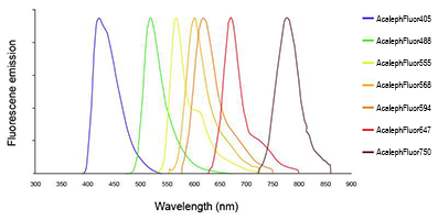 Line colors represent the approximate visible colors of the wavelength maxima.
 -
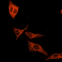 Immunofluorescent analysis of beta Tubulin staining in Hela cells. Formalin-fixed cells were permeabilized with 0.1% Triton X-100 in TBS for 5-10 minutes and blocked with 3% BSA-PBS for 30 minutes at room temperature. Cells were probed with the primary antibody in 3% BSA-PBS and incubated overnight at 4 °C in a hidified chamber. Cells were washed with PBST. The secondary antibody (orange) was Goat Anti-Rabbit IgG (H&L) - AF555 in PBS at room temperature in the dark.
Sunitinib-induced oxidative imbalance and retinotoxic effects in rats
Retinoprotective Effect of Wild Olive (Acebuche) Oil-Enriched Diet against Ocular Oxidative Stress Induced by Arterial Hypertension
NADPH oxidase–induced oxidative stress in the eyes of hypertensive rats
Hypertension secondary to nitric oxide depletion produces oxidative imbalance and inflammatory/fibrotic outcomes in the cornea of C57BL/6 mice