
 Anti-UBE2F Antibody
  Datasheet
 MSDS
MSDS
Description: Rabbit polyclonal antibody to UBE2F Immunogen: Recombinant full length protein of human UBE2F Purification: The antibody was purified by immunogen affinity chromatography. Clonality: Polyclonal Form: Liquid in 0.42% Potassium phosphate, 0.87% Sodium chloride, pH 7.3, 30% glycerol, and 0.01% sodium azide. Dilution: WB (1/500 - 1/2000), IH (1/50 - 1/200), IF/IC (1/50 - 1/200) Gene Symbol: UBE2F Alternative Names: NCE2; NEDD8-conjugating enzyme UBE2F; NEDD8 carrier protein UBE2F; NEDD8 protein ligase UBE2F; NEDD8-conjugating enzyme 2; Ubiquitin-conjugating enzyme E2 FEntrez Gene (Human): 140739Entrez Gene (Rat) : 363284SwissProt (Human): Q969M7SwissProt (Rat) : Q5U203Storage/Stability : Shipped at 4°C. Upon delivery aliquot and store at -20°C for one year. Avoid freeze/thaw cycles.
-
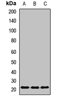 Western blot analysis of UBE2F expression in K562 (A), mouse liver (B), mouse heart (C) whole cell lysates.
 -
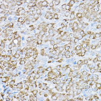 Immunohistochemical analysis of UBE2F staining in rat ovary formalin fixed paraffin embedded tissue section. The section was pre-treated using heat mediated antigen retrieval with sodium citrate buffer (pH 6.0). The section was then incubated with the antibody at room temperature and detected using an HRP conjugated compact polymer system. DAB was used as the chromogen. The section was then counterstained with haematoxylin and mounted with DPX.
 -
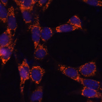 Immunofluorescent analysis of UBE2F staining in NIH3T3 cells. Formalin-fixed cells were permeabilized with 0.1% Triton X-100 in TBS for 5-10 minutes and blocked with 3% BSA-PBS for 30 minutes at room temperature. Cells were probed with the primary antibody in 3% BSA-PBS and incubated overnight at 4 °C in a humidified chamber. Cells were washed with PBST and incubated with a AF594-conjugated secondary antibody (red) in PBS at room temperature in the dark. DAPI was used to stain the cell nuclei (blue).