
 Anti-OB Cadherin Antibody
  Datasheet
 MSDS
MSDS
Description: Rabbit polyclonal antibody to OB Cadherin Immunogen: Recombinant full length protein of human OB Cadherin Purification: The antibody was purified by immunogen affinity chromatography. Clonality: Polyclonal Form: Liquid in 0.42% Potassium phosphate, 0.87% Sodium chloride, pH 7.3, 30% glycerol, and 0.01% sodium azide. Dilution: WB (1/500 - 1/1000), IF/IC (1/50 - 1/100) Gene Symbol: CDH11 Alternative Names: Cadherin-11; OSF-4; Osteoblast cadherin; OB-cadherinEntrez Gene (Human): 1009SwissProt (Human): P55287Storage/Stability : Shipped at 4°C. Upon delivery aliquot and store at -20°C for one year. Avoid freeze/thaw cycles.
-
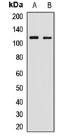 Western blot analysis of OB Cadherin expression in A549 (A), HT29 (B) whole cell lysates.
 -
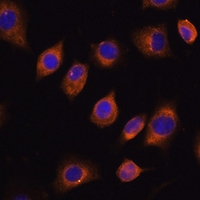 Immunofluorescent analysis of OB Cadherin staining in L929 cells. Formalin-fixed cells were permeabilized with 0.1% Triton X-100 in TBS for 5-10 minutes and blocked with 3% BSA-PBS for 30 minutes at room temperature. Cells were probed with the primary antibody in 3% BSA-PBS and incubated overnight at 4 °C in a humidified chamber. Cells were washed with PBST and incubated with a AF594-conjugated secondary antibody (red) in PBS at room temperature in the dark. DAPI was used to stain the cell nuclei (blue).