
 Anti-MATH-1 Antibody
  Datasheet
 MSDS
MSDS
Description: Rabbit polyclonal antibody to MATH-1 Immunogen: Recombinant full length protein of human MATH-1 Purification: The antibody was purified by immunogen affinity chromatography. Clonality: Polyclonal Form: Liquid in 0.42% Potassium phosphate, 0.87% Sodium chloride, pH 7.3, 30% glycerol, and 0.01% sodium azide. Dilution: WB (1/500 - 1/2000) Gene Symbol: ATOH1 Alternative Names: ATH1; BHLHA14; Protein atonal homolog 1; Class A basic helix-loop-helix protein 14; bHLHa14; Helix-loop-helix protein hATH-1; hATH1Entrez Gene (Human): 474SwissProt (Human): Q92858Storage/Stability : Shipped at 4°C. Upon delivery aliquot and store at -20°C for one year. Avoid freeze/thaw cycles.
-
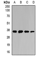 Western blot analysis of MATH-1 expression in SW620 (A), HepG2 (B), mouse liver (C), mouse heart (D) whole cell lysates.
 -
 Immunofluorescent analysis of MATH-1 staining in U2OS cells. Formalin-fixed cells were permeabilized with 0.1% Triton X-100 in TBS for 5-10 minutes and blocked with 3% BSA-PBS for 30 minutes at room temperature. Cells were probed with the primary antibody in 3% BSA-PBS and incubated overnight at 4 °C in a hidified chamber. Cells were washed with PBST and incubated with a DyLight 594-conjugated secondary antibody (red) in PBS at room temperature in the dark.