
 Anti-HADHB Antibody
  Datasheet
 MSDS
MSDS
Description: Rabbit polyclonal antibody to HADHB Immunogen: Recombinant full length protein of human HADHB Purification: The antibody was purified by immunogen affinity chromatography. Clonality: Polyclonal Form: Liquid in 0.42% Potassium phosphate, 0.87% Sodium chloride, pH 7.3, 30% glycerol, and 0.01% sodium azide. Dilution: WB (1/500 - 1/2000), IF/IC (1/10 - 1/100) Gene Symbol: HADHB Alternative Names: Trifunctional enzyme subunit beta mitochondrial; TP-betaEntrez Gene (Human): 3032Entrez Gene (Mouse) : 231086Entrez Gene (Rat) : 171155SwissProt (Human): P55084SwissProt (Mouse) : Q99JY0SwissProt (Rat) : Q60587Storage/Stability : Shipped at 4°C. Upon delivery aliquot and store at -20°C for one year. Avoid freeze/thaw cycles.
-
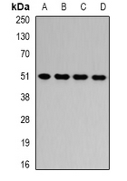 Western blot analysis of HADHB expression in HepG2 (A), Jurkat (B), mouse liver (C), rat liver (D) whole cell lysates.
 -
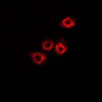 Immunofluorescent analysis of HADHB staining in A549 cells. Formalin-fixed cells were permeabilized with 0.1% Triton X-100 in TBS for 5-10 minutes and blocked with 3% BSA-PBS for 30 minutes at room temperature. Cells were probed with the primary antibody in 3% BSA-PBS and incubated overnight at 4 °C in a hidified chamber. Cells were washed with PBST and incubated with a DyLight 594-conjugated secondary antibody (red) in PBS at room temperature in the dark.