
 Anti-Cytokeratin 7 Antibody
  Datasheet
 MSDS
MSDS
Description: Mouse monoclonal antibody to Cytokeratin 7 Immunogen: KLH-conjugated synthetic peptide encompassing a sequence within human Cytokeratin 7. The exact sequence is proprietary. Purification: The antibody was purified by immunogen affinity chromatography. Clonality: Monoclonal Form: Mouse IgG2b. Liquid in PBS containing 50% glycerol, 0.2% BSA and 0.01% sodium azide. Dilution: WB (1/500 - 1/1000), IH (1/100 - 1/300) Gene Symbol: KRT7 Alternative Names: SCL; Keratin type II cytoskeletal 7; Cytokeratin-7; CK-7; Keratin-7; K7; Sarcolectin; Type-II keratin Kb7Entrez Gene (Human): 3855SwissProt (Human): P08729Storage/Stability : Shipped at 4°C. Upon delivery aliquot and store at -20°C for one year. Avoid freeze/thaw cycles.
-
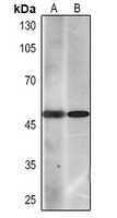 Western blot analysis of Cytokeratin 7 expression in Hela (A), A549 (B) whole cell lysates.
 -
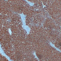 Immunohistochemical analysis of Cytokeratin 7 staining in human bladder transitional cell carcinoma formalin fixed paraffin embedded tissue section. The section was pre-treated using heat mediated antigen retrieval with sodium citrate buffer (pH 6.0). The section was then incubated with the antibody at room temperature and detected using an HRP conjugated compact polymer system. DAB was used as the chromogen. The section was then counterstained with haematoxylin and mounted with DPX.
 -
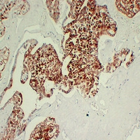 Immunohistochemical analysis of Cytokeratin 7 staining in human chromophobe cell renal carcinoma formalin fixed paraffin embedded tissue section. The section was pre-treated using heat mediated antigen retrieval with sodium citrate buffer (pH 6.0). The section was then incubated with the antibody at room temperature and detected using an HRP conjugated compact polymer system. DAB was used as the chromogen. The section was then counterstained with haematoxylin and mounted with DPX.
 -
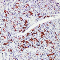
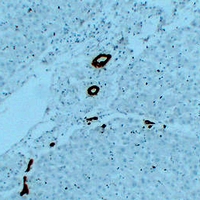 Immunohistochemical analysis of Cytokeratin 7 staining in human liver formalin fixed paraffin embedded tissue section. The section was pre-treated using heat mediated antigen retrieval with sodium citrate buffer (pH 6.0). The section was then incubated with the antibody at room temperature and detected using an HRP conjugated compact polymer system. DAB was used as the chromogen. The section was then counterstained with haematoxylin and mounted with DPX.
 -
 Immunohistochemical analysis of Cytokeratin 7 staining in human pancreas formalin fixed paraffin embedded tissue section. The section was pre-treated using heat mediated antigen retrieval with sodium citrate buffer (pH 6.0). The section was then incubated with the antibody at room temperature and detected using an HRP conjugated compact polymer system. DAB was used as the chromogen. The section was then counterstained with haematoxylin and mounted with DPX.