
 Anti-Methyl Lysine Antibody
  Datasheet
 MSDS
MSDS
Description: Mouse monoclonal antibody to Pan Methyl Lysine Immunogen: Purified protein corresponding to Methyl Lysine. Clonality: Monoclonal Form: Liquid in 0.42% Potassium phosphate, 0.87% Sodium chloride, pH 7.3, 30% glycerol, and 0.01% sodium azide. Dilution: WB (1/1000 - 1/2000), IH (1/200 - 1/500) Storage/Stability : Shipped at 4°C. Upon delivery aliquot and store at -20°C for one year. Avoid freeze/thaw cycles.
-
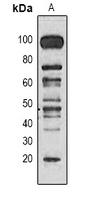 Western blot analysis of Methyl Lysine expression in Hela (A) whole cell lysates.
 -
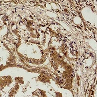 Immunohistochemical analysis of Methyl Lysine staining in human breast cancer formalin fixed paraffin embedded tissue section. The section was pre-treated using heat mediated antigen retrieval with sodium citrate buffer (pH 6.0). The section was then incubated with the antibody at room temperature and detected using an HRP conjugated compact polymer system. DAB was used as the chromogen. The section was then counterstained with haematoxylin and mounted with DPX.