
 Anti-Histone H3 Antibody
  Datasheet
 MSDS
MSDS
Description: Mouse monoclonal antibody to Histone H3 Immunogen: Recombinant protein corresponding to human Histone H3. Clonality: Monoclonal Form: Liquid in 0.42% Potassium phosphate, 0.87% Sodium chloride, pH 7.3, 30% glycerol, and 0.01% sodium azide. Dilution: WB (1/2000 - 1/5000), IF/IC (1/100 - 1/500), IP (1/50 - 1/200) Gene Symbol: HIST1H3A; HIST1H3B; HIST1H3C; HIST1H3D; HIST1H3E; HIST1H3F; HIST1H3G; HIST1H3H; HIST1H3I; HIST1H3J; HIST2H3A; HIST2H3C; HIST2H3D; H3F3A; H3F3B Alternative Names: HIST1H3A; H3FA; HIST1H3B; H3FL; HIST1H3C; H3FC; HIST1H3D; H3FB; HIST1H3E; H3FD; HIST1H3F; H3FI; HIST1H3G; H3FH; HIST1H3H; H3FK; HIST1H3I; H3FF; HIST1H3J; H3FJ; Histone H3.1; Histone H3/a; Histone H3/b; Histone H3/c; Histone H3/d; Histone H3/f; Histone H3/h; Histone H3/i; Histone H3/j; Histone H3/k; Histone H3/l; HIST2H3A; HIST2H3C; H3F2; H3FM; HIST2H3D; Histone H3.2; Histone H3/m; Histone H3/o; H3F3A; H3.3A; H3F3; PP781; H3F3B; H3.3B; Histone H3.3Entrez Gene (Human): 8350; 8351; 8352; 8353; 8354; 8355; 8356; 8357; 8358; 8968Entrez Gene (Mouse) : 319152; 15077; 15078Entrez Gene (Rat) : 291159; 100361558SwissProt (Human): P68431; Q71DI3; P84243SwissProt (Mouse) : P68433; P84228; P84244SwissProt (Rat) : Q6LED0; P84245Storage/Stability : Shipped at 4°C. Upon delivery aliquot and store at -20°C for one year. Avoid freeze/thaw cycles.
-
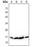 Western blot analysis of Histone H3 expression in Hela (A), Raw264.7 (B), mouse brain (C), rat brain (D) whole cell lysates.
 -
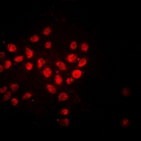 Immunofluorescent analysis of Histone H3 staining in Hela cells. Formalin-fixed cells were permeabilized with 0.1% Triton X-100 in TBS for 5-10 minutes and blocked with 3% BSA-PBS for 30 minutes at room temperature. Cells were probed with the primary antibody in 3% BSA-PBS and incubated overnight at 4 °C in a hidified chamber. Cells were washed with PBST and incubated with a DyLight 594-conjugated secondary antibody (red) in PBS at room temperature in the dark.
 -
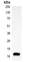 Immunoprecipitation of Histone H3 from 0.5mg Hela whole cell extract lysate, using Anti-Histone H3 Antibody.