
 Anti-Alpha-tubulin Antibody
  Datasheet
 MSDS
MSDS
Description: Mouse monoclonal antibody to Alpha-tubulin Immunogen: Recombinant protein corresponding to human Alpha-tubulin. Clonality: Monoclonal Form: Liquid in 0.42% Potassium phosphate, 0.87% Sodium chloride, pH 7.3, 0.2% BSA, 30% glycerol, and 0.01% sodium azide. Dilution: WB (1/3000 - 1/10000), IH (1/50 - 1/100), IF/IC (1/50 - 1/100), IP (1/50 - 1/100) Gene Symbol: TUBA1A Alternative Names: TUBA3; Tubulin alpha-1A chain; Alpha-tubulin 3; Tubulin B-alpha-1; Tubulin alpha-3 chainEntrez Gene (Human): 7846; 10376Entrez Gene (Mouse) : 22142; 22143Entrez Gene (Rat) : 64158; 500929SwissProt (Human): Q71U36; P68363SwissProt (Mouse) : P68369; P05213SwissProt (Rat) : P68370; Q6P9V9Storage/Stability : Shipped at 4°C. Upon delivery aliquot and store at -20°C for one year. Avoid freeze/thaw cycles.
-
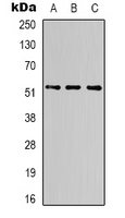 Western blot analysis of Alpha-tubulin expression in Hela (A), mouse brain (B), rat brain (C) whole cell lysates.
 -
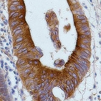 Immunohistochemical analysis of Alpha-tubulin staining in human colon cancer formalin fixed paraffin embedded tissue section. The section was pre-treated using heat mediated antigen retrieval with sodium citrate buffer (pH 6.0). The section was then incubated with the antibody at room temperature and detected using an HRP conjugated compact polymer system. DAB was used as the chromogen. The section was then counterstained with haematoxylin and mounted with DPX.
 -
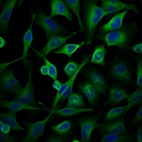 Immunofluorescent analysis of Alpha-tubulin staining in Hela cells. Formalin-fixed cells were permeabilized with 0.1% Triton X-100 in TBS for 5-10 minutes and blocked with 3% BSA-PBS for 30 minutes at room temperature. Cells were probed with the primary antibody in 3% BSA-PBS and incubated overnight at 4 °C in a hidified chamber. Cells were washed with PBST and incubated with a FITC-conjugated secondary antibody (green) in PBS at room temperature in the dark. DAPI was used to stain the cell nuclei (blue).
 -
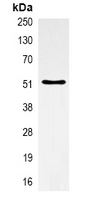 Immunoprecipitation of Alpha-tubulin from 0.5mg mouse brain whole cell extract lysate, using Anti-Alpha-tubulin Antibody.
Hericium erinaceus, in combination with natural flavonoid/alkaloid and B3/B8 vitamins, can improve inflammatory burden in Inflammatory bowel diseases tissue: an ex vivo study