
 Anti-p38 (Phospho-Y182) Antibody
  Datasheet
 MSDS
MSDS
Description: Rabbit polyclonal antibody to p38 (Phospho-Y182) Immunogen: KLH-conjugated synthetic phosphopeptide corresponding to residues surrounding Y182 of human p38 protein. The exact sequence is proprietary. Purification: The antibody was purified by immunogen affinity chromatography. Clonality: Polyclonal Form: Liquid in 0.42% Potassium phosphate, 0.87% Sodium chloride, pH 7.3, 30% glycerol, and 0.01% sodium azide. Dilution: WB (1/500 - 1/1000), IH (1/100 - 1/200), IF/IC (1/100 - 1/500) Gene Symbol: MAPK14 Alternative Names: CSBP; CSBP1; CSBP2; CSPB1; MXI2; SAPK2A; Mitogen-activated protein kinase 14; MAP kinase 14; MAPK 14; Cytokine suppressive anti-inflammatory drug-binding protein; CSAID-binding protein; CSBP; MAP kinase MXI2; MAX-interacting protein 2; Mitogen-activated protein kinase p38 alpha; MAP kinase p38 alpha; Stress-activated protein kinase 2a; SAPK2aEntrez Gene (Human): 1432Entrez Gene (Mouse) : 26416SwissProt (Human): Q16539SwissProt (Mouse) : P47811SwissProt (Rat) : P70618Storage/Stability : Shipped at 4°C. Upon delivery aliquot and store at -20°C for one year. Avoid freeze/thaw cycles.
-
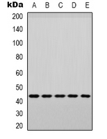 Western blot analysis of p38 (Phospho-Y182) expression in PC3 (A), MCF7 (B), Raw264.7 (C), mouse muscle (D), rat muscle (E) whole cell lysates.
 -
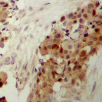 Immunohistochemical analysis of p38 (Phospho-Y182) staining in human breast cancer formalin fixed paraffin embedded tissue section. The section was pre-treated using heat mediated antigen retrieval with sodium citrate buffer (pH 6.0). The section was then incubated with the antibody at room temperature and detected using an HRP conjugated compact polymer system. DAB was used as the chromogen. The section was then counterstained with haematoxylin and mounted with DPX.
 -
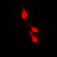 Immunofluorescent analysis of p38 (Phospho-Y182) staining in HepG2 cells. Formalin-fixed cells were permeabilized with 0.1% Triton X-100 in TBS for 5-10 minutes and blocked with 3% BSA-PBS for 30 minutes at room temperature. Cells were probed with the primary antibody in 3% BSA-PBS and incubated overnight at 4 °C in a hidified chamber. Cells were washed with PBST and incubated with a DyLight 594-conjugated secondary antibody (red) in PBS at room temperature in the dark.
 -
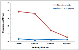 Direct ELISA antibody dose-response curve using Anti-p38 (Phospho-Y182) Antibody. Antigen (Phosphopeptide and non-phosphopeptide) concentration is 5 ug/ml. Goat Anti-Rabbit IgG (H&L) - HRP was used as the secondary antibody, and signal was developed by TMB substrate.