
 Anti-CD4 (Phospho-S433) Antibody
  Datasheet
 MSDS
MSDS
Description: Rabbit polyclonal antibody to CD4 (Phospho-S433) Immunogen: KLH-conjugated synthetic phosphopeptide corresponding to residues surrounding S433 of human CD4 protein. The exact sequence is proprietary. Purification: The antibody was purified by immunogen affinity chromatography. Clonality: Polyclonal Form: Liquid in 0.42% Potassium phosphate, 0.87% Sodium chloride, pH 7.3, 30% glycerol, and 0.01% sodium azide. Dilution: WB (1/500 - 1/1000), IH (1/50 - 1/200), IF/IC (1/50 - 1/200) Gene Symbol: CD4 Alternative Names: T-cell surface glycoprotein CD4; T-cell surface antigen T4/Leu-3; CD4Entrez Gene (Human): 920Entrez Gene (Mouse) : 12504Entrez Gene (Rat) : 24932SwissProt (Human): P01730SwissProt (Mouse) : P06332SwissProt (Rat) : P05540Storage/Stability : Shipped at 4°C. Upon delivery aliquot and store at -20°C for one year. Avoid freeze/thaw cycles.
-
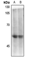 Western blot analysis of CD4 (pS433) expression in mouse spleen (A), rat spleen (B) whole cell lysates.
 -
 Immunofluorescent analysis of CD4 (Phospho-S433) staining in HepG2 cells. Formalin-fixed cells were permeabilized with 0.1% Triton X-100 in TBS for 5-10 minutes and blocked with 3% BSA-PBS for 30 minutes at room temperature. Cells were probed with the primary antibody in 3% BSA-PBS and incubated overnight at 4 °C in a hidified chamber. Cells were washed with PBST and incubated with a Alexa Fluor 594-conjugated secondary antibody (red) in PBS at room temperature in the dark.
 -
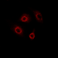 Immunofluorescent analysis of CD4 (pS433) staining in HepG2 cells. Formalin-fixed cells were permeabilized with 0.1% Triton X-100 in TBS for 5-10 minutes and blocked with 3% BSA-PBS for 30 minutes at room temperature. Cells were probed with the primary antibody in 3% BSA-PBS and incubated overnight at 4 °C in a hidified chamber. Cells were washed with PBST and incubated with a Alexa Fluor 594-conjugated secondary antibody (red) in PBS at room temperature in the dark.
 -
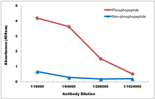 Direct ELISA antibody dose-response curve using Anti-CD4 (Phospho-S433) Antibody. Antigen (Phosphopeptide and non-phosphopeptide) concentration is 5 ug/ml. Goat Anti-Rabbit IgG (H&L) - HRP was used as the secondary antibody, and signal was developed by TMB substrate.