
 Anti-AS160 Antibody
  Datasheet
 MSDS
MSDS
Description: Rabbit polyclonal antibody to AS160 Immunogen: KLH-conjugated synthetic peptide encompassing a sequence within the center region of human AS160. The exact sequence is proprietary. Purification: The antibody was purified by immunogen affinity chromatography. Clonality: Polyclonal Form: Liquid in 0.42% Potassium phosphate, 0.87% Sodium chloride, pH 7.3, 30% glycerol, and 0.01% sodium azide. Dilution: WB (1/500 - 1/1000), IH (1/100 - 1/200), IF/IC (1/100 - 1/500) Gene Symbol: TBC1D4 Alternative Names: AS160; KIAA0603; TBC1 domain family member 4; Akt substrate of 160 kDa; AS160Entrez Gene (Human): 9882Entrez Gene (Mouse) : 210789SwissProt (Human): O60343SwissProt (Mouse) : Q8BYJ6Storage/Stability : Shipped at 4°C. Upon delivery aliquot and store at -20°C for one year. Avoid freeze/thaw cycles.
-
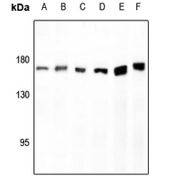 Western blot analysis of AS160 expression in H9C2 (A), U87MG (B), HUT78 (C), Myla2059 (D), HepG2 (E), HEK293T (F) whole cell lysates.
 -
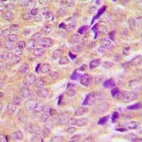 Immunohistochemical analysis of AS160 staining in human breast cancer formalin fixed paraffin embedded tissue section. The section was pre-treated using heat mediated antigen retrieval with sodium citrate buffer (pH 6.0). The section was then incubated with the antibody at room temperature and detected using an HRP conjugated compact polymer system. DAB was used as the chromogen. The section was then counterstained with haematoxylin and mounted with DPX.
 -
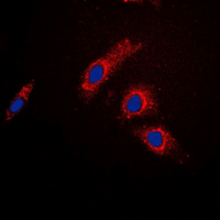 Immunofluorescent analysis of AS160 staining in HeLa cells. Formalin-fixed cells were permeabilized with 0.1% Triton X-100 in TBS for 5-10 minutes and blocked with 3% BSA-PBS for 30 minutes at room temperature. Cells were probed with the primary antibody in 3% BSA-PBS and incubated overnight at 4 °C in a hidified chamber. Cells were washed with PBST and incubated with a DyLight 594-conjugated secondary antibody (red) in PBS at room temperature in the dark. DAPI was used to stain the cell nuclei (blue).