
 Anti-DARPP32 (Phospho-T34) Antibody
  Datasheet
 MSDS
MSDS
Description: Rabbit polyclonal antibody to DARPP32 (Phospho-T34) Immunogen: KLH-conjugated synthetic phosphopeptide corresponding to residues surrounding T34 of human DARPP32 protein. The exact sequence is proprietary. Purification: The antibody was purified by immunogen affinity chromatography. Clonality: Polyclonal Form: Liquid in 0.42% Potassium phosphate, 0.87% Sodium chloride, pH 7.3, 30% glycerol, and 0.01% sodium azide. Dilution: WB (1/500 - 1/1000), IH (1/100 - 1/200), IF/IC (1/100 - 1/500) Gene Symbol: PPP1R1B Alternative Names: DARPP32; Protein phosphatase 1 regulatory subunit 1B; DARPP-32; Dopamine- and cAMP-regulated neuronal phosphoproteinEntrez Gene (Human): 84152Entrez Gene (Mouse) : 19049SwissProt (Human): Q9UD71SwissProt (Mouse) : Q60829Storage/Stability : Shipped at 4°C. Upon delivery aliquot and store at -20°C for one year. Avoid freeze/thaw cycles.
-
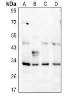 Western blot analysis of DARPP32 (Phospho-T34) expression in HEK293T (A), HepG2 (B), MCF7 (C), mouse spleen (D) whole cell lysates.
 -
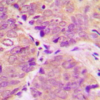 Immunohistochemical analysis of DARPP32 (Phospho-T34) staining in human breast cancer formalin fixed paraffin embedded tissue section. The section was pre-treated using heat mediated antigen retrieval with sodium citrate buffer (pH 6.0). The section was then incubated with the antibody at room temperature and detected using an HRP conjugated compact polymer system. DAB was used as the chromogen. The section was then counterstained with haematoxylin and mounted with DPX.
 -
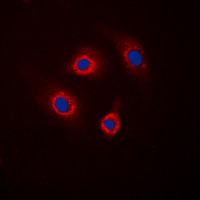 Immunofluorescent analysis of DARPP32 (Phospho-T34) staining in HepG2 cells. Formalin-fixed cells were permeabilized with 0.1% Triton X-100 in TBS for 5-10 minutes and blocked with 3% BSA-PBS for 30 minutes at room temperature. Cells were probed with the primary antibody in 3% BSA-PBS and incubated overnight at 4 °C in a hidified chamber. Cells were washed with PBST and incubated with a DyLight 594-conjugated secondary antibody (red) in PBS at room temperature in the dark. DAPI was used to stain the cell nuclei (blue).
 -
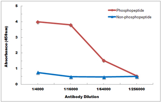 Direct ELISA antibody dose-response curve using Anti-DARPP32 (Phospho-T34) Antibody. Antigen (Phosphopeptide and non-phosphopeptide) concentration is 5 ug/ml. Goat Anti-Rabbit IgG (H&L) - HRP was used as the secondary antibody, and signal was developed by TMB substrate.