
 Anti-c-Met (Phospho-Y1349) Antibody
  Datasheet
 MSDS
MSDS
Description: Rabbit polyclonal antibody to c-Met (Phospho-Y1349) Immunogen: KLH-conjugated synthetic phosphopeptide corresponding to residues surrounding Y1349 of human c-Met protein. The exact sequence is proprietary. Purification: The antibody was purified by immunogen affinity chromatography. Clonality: Polyclonal Form: Liquid in 0.42% Potassium phosphate, 0.87% Sodium chloride, pH 7.3, 30% glycerol, and 0.01% sodium azide. Dilution: WB (1/500 - 1/1000), IH (1/50 - 1/100), IF/IC (1/50 - 1/200) Gene Symbol: MET Alternative Names: Hepatocyte growth factor receptor; HGF receptor; HGF/SF receptor; Proto-oncogene c-Met; Scatter factor receptor; SF receptor; Tyrosine-protein kinase MetEntrez Gene (Human): 4233Entrez Gene (Rat) : 24553SwissProt (Human): P08581SwissProt (Mouse) : P16056SwissProt (Rat) : P97523Storage/Stability : Shipped at 4°C. Upon delivery aliquot and store at -20°C for one year. Avoid freeze/thaw cycles.
-
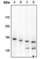 Western blot analysis of c-Met (Phospho-Y1349) expression in CT26 (A), rat liver (B), HEK293T (C), LO2 (D) whole cell lysates.
 -
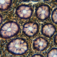 Immunohistochemical analysis of c-Met (Phospho-Y1349) staining in human colorectal cancer formalin fixed paraffin embedded tissue section. The section was pre-treated using heat mediated antigen retrieval with sodium citrate buffer (pH 6.0). The section was then incubated with the antibody at room temperature and detected using an HRP conjugated compact polymer system. DAB was used as the chromogen. The section was then counterstained with haematoxylin and mounted with DPX.
 -
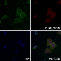 Immunofluorescent analysis of c-Met (Phospho-Y1349) staining in LO2 cells. Formalin-fixed cells were permeabilized with 0.1% Triton X-100 in TBS for 5-10 minutes and blocked with 3% BSA-PBS for 30 minutes at room temperature. Cells were probed with the primary antibody in 3% BSA-PBS and incubated overnight at 4 °C in a hidified chamber. Cells were washed with PBST and incubated with a AF488-conjugated secondary antibody (green) in PBS at room temperature in the dark. Phalloidin - AF594 was used to stain Actin filaments (red). DAPI was used to stain the cell nuclei (blue).
Development of a cell-based assay for measurement of c-Met phosphorylation using AlphaScreen technology and high-content imaging analysis