
 Anti-Aurora A (Phospho-T288) Antibody
  Datasheet
 MSDS
MSDS
Description: Rabbit polyclonal antibody to Aurora A (Phospho-T288) Immunogen: KLH-conjugated synthetic phosphopeptide corresponding to residues surrounding T288 of human Aurora A protein. The exact sequence is proprietary. Purification: The antibody was purified by immunogen affinity chromatography. Clonality: Polyclonal Form: Liquid in 0.42% Potassium phosphate, 0.87% Sodium chloride, pH 7.3, 30% glycerol, and 0.01% sodium azide. Dilution: WB (1/500 - 1/1000), IH (1/100 - 1/200), IF/IC (1/100 - 1/500) Gene Symbol: AURKA Alternative Names: AIK; AIRK1; ARK1; AURA; AYK1; BTAK; IAK1; STK15; STK6; Aurora kinase A; Aurora 2; Aurora/IPL1-related kinase 1; ARK-1; Aurora-related kinase 1; hARK1; Breast tumor-amplified kinase; Serine/threonine-protein kinase 15; Serine/threonine-protein kinase 6; Serine/threonine-protein kinase aurora-AEntrez Gene (Human): 6790Entrez Gene (Mouse) : 20878SwissProt (Human): O14965SwissProt (Mouse) : P97477SwissProt (Rat) : P59241Storage/Stability : Shipped at 4°C. Upon delivery aliquot and store at -20°C for one year. Avoid freeze/thaw cycles.
-
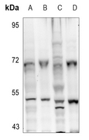 Western blot analysis of Aurora A (Phospho-T288) expression in HCT116 (A), SHSY5Y (B), CT26 (C), C6 (D) whole cell lysates.
 -
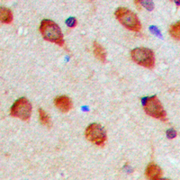 Immunohistochemical analysis of Aurora A (Phospho-T288) staining in human brain formalin fixed paraffin embedded tissue section. The section was pre-treated using heat mediated antigen retrieval with sodium citrate buffer (pH 6.0). The section was then incubated with the antibody at room temperature and detected using an HRP conjugated compact polymer system. DAB was used as the chromogen. The section was then counterstained with haematoxylin and mounted with DPX.
 -
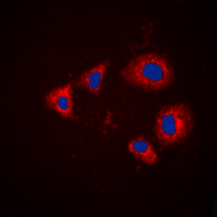 Immunofluorescent analysis of Aurora A (Phospho-T288) staining in HEK293T cells. Formalin-fixed cells were permeabilized with 0.1% Triton X-100 in TBS for 5-10 minutes and blocked with 3% BSA-PBS for 30 minutes at room temperature. Cells were probed with the primary antibody in 3% BSA-PBS and incubated overnight at 4 °C in a hidified chamber. Cells were washed with PBST and incubated with a DyLight 594-conjugated secondary antibody (red) in PBS at room temperature in the dark. DAPI was used to stain the cell nuclei (blue).
Differential expression of aurora-A kinase in T-cell lymphomas
Relationship of increased aurora kinase A gene copy number, prognosis and response to chemotherapy in patients with metastatic colorectal cancer
Role of Filia, a maternal effect gene, in maintaining euploidy during cleavage-stage mouse embryogenesis