
 Anti-ATP5L2 Antibody
  Datasheet
 MSDS
MSDS
Description: Rabbit polyclonal antibody to ATP5L2 Immunogen: KLH-conjugated synthetic peptide encompassing a sequence within the center region of human ATP5L2. The exact sequence is proprietary. Purification: The antibody was purified by immunogen affinity chromatography. Clonality: Polyclonal Form: Liquid in 0.42% Potassium phosphate, 0.87% Sodium chloride, pH 7.3, 30% glycerol, and 0.01% sodium azide. Dilution: WB (1/500 - 1/1000), IF/IC (1/100 - 1/500) Gene Symbol: ATP5L2 Alternative Names: ATP5K2; ATP synthase subunit g 2 mitochondrial; ATPase subunit g 2Entrez Gene (Human): 267020SwissProt (Human): Q7Z4Y8Storage/Stability : Shipped at 4°C. Upon delivery aliquot and store at -20°C for one year. Avoid freeze/thaw cycles.
-
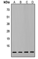 Western blot analysis of ATP5L2 expression in HepG2 (A), PC3 (B), MCF7 (C), NIH3T3 (D) whole cell lysates.
 -
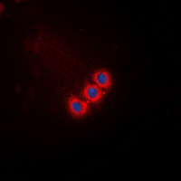 Immunofluorescent analysis of ATP5L2 staining in HepG2 cells. Formalin-fixed cells were permeabilized with 0.1% Triton X-100 in TBS for 5-10 minutes and blocked with 3% BSA-PBS for 30 minutes at room temperature. Cells were probed with the primary antibody in 3% BSA-PBS and incubated overnight at 4 °C in a hidified chamber. Cells were washed with PBST and incubated with a DyLight 594-conjugated secondary antibody (red) in PBS at room temperature in the dark. DAPI was used to stain the cell nuclei (blue).