
 Anti-VDR (Phospho-S208) Antibody
  Datasheet
 MSDS
MSDS
Description: Rabbit polyclonal antibody to VDR (Phospho-S208) Immunogen: KLH-conjugated synthetic phosphopeptide corresponding to residues surrounding S208 of human VDR protein. The exact sequence is proprietary. Purification: The antibody was purified by immunogen affinity chromatography. Clonality: Polyclonal Form: Liquid in 0.42% Potassium phosphate, 0.87% Sodium chloride, pH 7.3, 30% glycerol, and 0.01% sodium azide. Dilution: WB (1/500 - 1/1000), IF/IC (1/100 - 1/500) Gene Symbol: VDR Alternative Names: NR1I1; Vitamin D3 receptor; VDR; 1,25-dihydroxyvitamin D3 receptor; Nuclear receptor subfamily 1 group I member 1Entrez Gene (Human): 7421Entrez Gene (Mouse) : 22337Entrez Gene (Rat) : 24873SwissProt (Human): P11473SwissProt (Mouse) : P48281SwissProt (Rat) : P13053Storage/Stability : Shipped at 4°C. Upon delivery aliquot and store at -20°C for one year. Avoid freeze/thaw cycles.
-
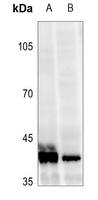 Western blot analysis of VDR (Phospho-S208) expression in HCT116 (A), HEK293T (B) whole cell lysates.
 -
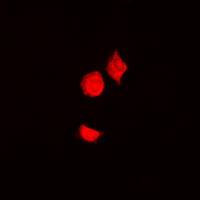 Immunofluorescent analysis of VDR (Phospho-S208) staining in HepG2 cells. Formalin-fixed cells were permeabilized with 0.1% Triton X-100 in TBS for 5-10 minutes and blocked with 3% BSA-PBS for 30 minutes at room temperature. Cells were probed with the primary antibody in 3% BSA-PBS and incubated overnight at 4 °C in a hidified chamber. Cells were washed with PBST and incubated with a DyLight 594-conjugated secondary antibody (red) in PBS at room temperature in the dark. DAPI was used to stain the cell nuclei (blue).
Targeting iron homeostasis induces cellular differentiation and synergizes with differentiating agents in acute myeloid leukemia