
 Anti-SP1 (Phospho-T453) Antibody
  Datasheet
 MSDS
MSDS
Description: Rabbit polyclonal antibody to SP1 (Phospho-T453) Immunogen: KLH-conjugated synthetic phosphopeptide corresponding to residues surrounding T453 of human SP1 protein. The exact sequence is proprietary. Purification: The antibody was purified by immunogen affinity chromatography. Clonality: Polyclonal Form: Liquid in 0.42% Potassium phosphate, 0.87% Sodium chloride, pH 7.3, 30% glycerol, and 0.01% sodium azide. Dilution: WB (1/500 - 1/1000), IH (1/100 - 1/200), IF/IC (1/100 - 1/500), ChIP (Use at an assay dependent concentration) Gene Symbol: SP1 Alternative Names: TSFP1; Transcription factor Sp1Entrez Gene (Human): 6667Entrez Gene (Mouse) : 20683Entrez Gene (Rat) : 24790SwissProt (Human): P08047SwissProt (Mouse) : O89090SwissProt (Rat) : Q01714Storage/Stability : Shipped at 4°C. Upon delivery aliquot and store at -20°C for one year. Avoid freeze/thaw cycles.
-
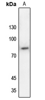 Western blot analysis of SP1 (Phospho-T453) expression in H1688 (A) whole cell lysates.
 -
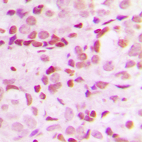 Immunohistochemical analysis of SP1 (Phospho-T453) staining in human breast cancer formalin fixed paraffin embedded tissue section. The section was pre-treated using heat mediated antigen retrieval with sodium citrate buffer (pH 6.0). The section was then incubated with the antibody at room temperature and detected using an HRP conjugated compact polymer system. DAB was used as the chromogen. The section was then counterstained with haematoxylin and mounted with DPX.
 -
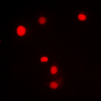 Immunofluorescent analysis of SP1 (Phospho-T453) staining in Jurkat cells. Formalin-fixed cells were permeabilized with 0.1% Triton X-100 in TBS for 5-10 minutes and blocked with 3% BSA-PBS for 30 minutes at room temperature. Cells were probed with the primary antibody in 3% BSA-PBS and incubated overnight at 4 °C in a hidified chamber. Cells were washed with PBST and incubated with a DyLight 594-conjugated secondary antibody (red) in PBS at room temperature in the dark.
A molecular prognostic model predicts esophageal squamous cell carcinoma prognosis
Multiple E-boxes in the distal promoter of the rat pyruvate carboxylase gene function as a glucose-responsive element
Dephosphorylation of Sp1 at Ser-59 by protein phosphatase 2A (PP2A) is required for induction of CYP1A1 transcription after treatment with 2,3,7,8-tetrachlorodibenzo-p-dioxin or omeprazole
NF-kB and enhancer-binding CREB protein scaffolded by CREB-binding protein (CBP)/p300 proteins regulate CD59 protein expression to protect cells from complement attack
Calcium input potentiates the transforming growth factor (TGF)-beta1-dependent signaling to promote the export of inorganic pyrophosphate by articular chondrocyte
The homeobox gene MSX2 determines chemosensitivity of pancreatic cancer cells via the regulation of transporter gene ABCG2
Heme Oxygenase-1/Carbon Monoxide Induces Vascular Endothelial Growth Factor Expression via p38 Kinase-dependent Activation of Sp1
Rac1-mediated Mitochondrial H2O2 Generation Regulates MMP-9 Gene Expression in Macrophages via Inhibition of SP-1 and AP-1
Regulation of embryonic kidney branching morphogenesis and glomerular development by KISS1 receptor (Gpr54) through NFAT2- and Sp1-mediated Bmp7 expression
Activated protein C therapy slows ALS-like disease in mice by transcriptionally inhibiting SOD1 in motor neurons and microglia cells
Hepatitis C virus core protein triggers hepatic angiogenesis by a mechanism including multiple pathways
The role of Sp1 in IL-1beta and H. pylori-mediated regulation of H,K-ATPase gene transcription