
 Anti-SMAD2 (Phospho-S467) Antibody
  Datasheet
 MSDS
MSDS
Description: Rabbit polyclonal antibody to SMAD2 (Phospho-S467) Immunogen: KLH-conjugated synthetic phosphopeptide corresponding to residues surrounding S467 of human SMAD2 protein. The exact sequence is proprietary. Purification: The antibody was purified by immunogen affinity chromatography. Clonality: Polyclonal Form: Liquid in 0.42% Potassium phosphate, 0.87% Sodium chloride, pH 7.3, 30% glycerol, and 0.01% sodium azide. Dilution: E (1/5000 - 1/10000), WB (1/500 - 1/1000), IH (1/100 - 1/200), IF/IC (1/100 - 1/500) Gene Symbol: SMAD2 Alternative Names: MADH2; MADR2; Mothers against decapentaplegic homolog 2; MAD homolog 2; Mothers against DPP homolog 2; JV18-1; Mad-related protein 2; hMAD-2; SMAD family member 2; SMAD 2; Smad2; hSMAD2Entrez Gene (Human): 4087Entrez Gene (Mouse) : 17126Entrez Gene (Rat) : 29357SwissProt (Human): Q15796SwissProt (Mouse) : Q62432SwissProt (Rat) : O70436Storage/Stability : Shipped at 4°C. Upon delivery aliquot and store at -20°C for one year. Avoid freeze/thaw cycles.
-
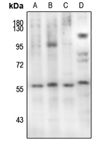 Western blot analysis of SMAD2 (Phospho-S467) expression in MCF7 (A), A375 (B), A549 (C), rat heart (D) whole cell lysates.
 -
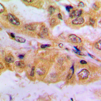 Immunohistochemical analysis of SMAD2 (Phospho-S467) staining in human lung cancer formalin fixed paraffin embedded tissue section. The section was pre-treated using heat mediated antigen retrieval with sodium citrate buffer (pH 6.0). The section was then incubated with the antibody at room temperature and detected using an HRP conjugated compact polymer system. DAB was used as the chromogen. The section was then counterstained with haematoxylin and mounted with DPX.
 -
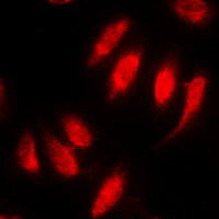 Immunofluorescent analysis of SMAD2 (Phospho-S467) staining in HeLa cells. Formalin-fixed cells were permeabilized with 0.1% Triton X-100 in TBS for 5-10 minutes and blocked with 3% BSA-PBS for 30 minutes at room temperature. Cells were probed with the primary antibody in 3% BSA-PBS and incubated overnight at 4 °C in a hidified chamber. Cells were washed with PBST and incubated with a DyLight 594-conjugated secondary antibody (red) in PBS at room temperature in the dark.
 -
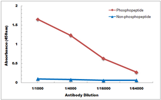 Direct ELISA antibody dose-response curve using Anti-SMAD2 (Phospho-S467) Antibody. Antigen (Phosphopeptide and non-phosphopeptide) concentration is 5 ug/ml. Goat Anti-Rabbit IgG (H&L) - HRP was used as the secondary antibody, and signal was developed by TMB substrate.
Matrix metalloproteinase-9 deletion attenuates myocardial fibrosis and diastolic dysfunction in ageing mice
Bone resorption and remodeling in murine collagenase-induced osteoarthritis after administration of glucosamine
A novel glycosylation signal regulates transforming growth factor beta receptors as evidenced by endo-beta-galactosidase C expression in rodent cells
Transforming growth factor-beta1 attenuates expression of both the progesterone receptor and Dickkopf in differentiated human endometrial stromal cells