
 Anti-p47 phox (Phospho-S359) Antibody
  Datasheet
 MSDS
MSDS
Description: Rabbit polyclonal antibody to p47 phox (Phospho-S359) Immunogen: KLH-conjugated synthetic phosphopeptide corresponding to residues surrounding S359 of human p47 phox protein. The exact sequence is proprietary. Purification: The antibody was purified by immunogen affinity chromatography. Clonality: Polyclonal Form: Liquid in 0.42% Potassium phosphate, 0.87% Sodium chloride, pH 7.3, 30% glycerol, and 0.01% sodium azide. Dilution: WB (1/500 - 1/1000), IH (1/100 - 1/200), IF/IC (1/100 - 1/500) Gene Symbol: NCF1 Alternative Names: NOXO2; SH3PXD1A; Neutrophil cytosol factor 1; NCF-1; 47 kDa autosomal chronic granulomatous disease protein; 47 kDa neutrophil oxidase factor; NCF-47K; Neutrophil NADPH oxidase factor 1; Nox organizer 2; Nox-organizing protein 2; SH3 and PX domain-containing protein 1A; p47-phoxEntrez Gene (Human): 653361SwissProt (Human): P14598Storage/Stability : Shipped at 4°C. Upon delivery aliquot and store at -20°C for one year. Avoid freeze/thaw cycles.
-
 Western blot analysis of p47 phox (Phospho-S359) expression in HeLa (A) whole cell lysates.
 -
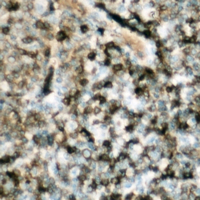 Immunohistochemical analysis of p47 phox (Phospho-S359) staining in human lymph node formalin fixed paraffin embedded tissue section. The section was pre-treated using heat mediated antigen retrieval with sodium citrate buffer (pH 6.0). The section was then incubated with the antibody at room temperature and detected using an HRP conjugated compact polymer system. DAB was used as the chromogen. The section was then counterstained with haematoxylin and mounted with DPX.
 -
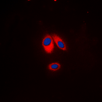 Immunofluorescent analysis of p47 phox (Phospho-S359) staining in HeLa cells. Formalin-fixed cells were permeabilized with 0.1% Triton X-100 in TBS for 5-10 minutes and blocked with 3% BSA-PBS for 30 minutes at room temperature. Cells were probed with the primary antibody in 3% BSA-PBS and incubated overnight at 4 °C in a hidified chamber. Cells were washed with PBST and incubated with a DyLight 594-conjugated secondary antibody (red) in PBS at room temperature in the dark. DAPI was used to stain the cell nuclei (blue).
NADPH oxidase expression in active multiple sclerosis lesions in relation to oxidative tissue damage and mitochondrial injury