
 Anti-EEF2 (Phospho-T56) Antibody
  Datasheet
 MSDS
MSDS
Description: Rabbit polyclonal antibody to EEF2 (Phospho-T56) Immunogen: KLH-conjugated synthetic phosphopeptide corresponding to residues surrounding T56 of human EEF2 protein. The exact sequence is proprietary. Purification: The antibody was purified by immunogen affinity chromatography. Clonality: Polyclonal Form: Liquid in 0.42% Potassium phosphate, 0.87% Sodium chloride, pH 7.3, 30% glycerol, and 0.01% sodium azide. Dilution: WB (1/500 - 1/1000), IH (1/100 - 1/200), IF/IC (1/100 - 1/500) Gene Symbol: EEF2 Alternative Names: EF2; Elongation factor 2; EF-2Entrez Gene (Human): 1938Entrez Gene (Mouse) : 13629Entrez Gene (Rat) : 29565SwissProt (Human): P13639SwissProt (Mouse) : P58252SwissProt (Rat) : P05197Storage/Stability : Shipped at 4°C. Upon delivery aliquot and store at -20°C for one year. Avoid freeze/thaw cycles.
-
 Western blot analysis of EEF2 (Phospho-T56) expression in DLD (A), U2OS (B) whole cell lysates.
 -
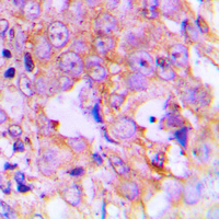 Immunohistochemical analysis of EEF2 (Phospho-T56) staining in human lung cancer formalin fixed paraffin embedded tissue section. The section was pre-treated using heat mediated antigen retrieval with sodium citrate buffer (pH 6.0). The section was then incubated with the antibody at room temperature and detected using an HRP conjugated compact polymer system. DAB was used as the chromogen. The section was then counterstained with haematoxylin and mounted with DPX.
 -
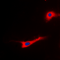 Immunofluorescent analysis of EEF2 (Phospho-T56) staining in SKOV3 cells. Formalin-fixed cells were permeabilized with 0.1% Triton X-100 in TBS for 5-10 minutes and blocked with 3% BSA-PBS for 30 minutes at room temperature. Cells were probed with the primary antibody in 3% BSA-PBS and incubated overnight at 4 °C in a hidified chamber. Cells were washed with PBST and incubated with a DyLight 594-conjugated secondary antibody (red) in PBS at room temperature in the dark. DAPI was used to stain the cell nuclei (blue).
Hypoxia Integration in the Serological Proteome Analysis Unmasks Tumor Antigens and Fosters the Identification of Anti-Phospho-eEF2 Antibodies as Potential Cancer Biomarkers
Identification and quantification of concentration-dependent biomarkers in MCF-7/BOS cells exposed to 17ß-estradiol by 2-D DIGE and label-free proteomics