
 Anti-Claudin 3 Antibody
  Datasheet
 MSDS
MSDS
Description: Rabbit polyclonal antibody to Claudin 3 Immunogen: KLH-conjugated synthetic peptide encompassing a sequence within the C-term region of human Claudin 3. The exact sequence is proprietary. Purification: The antibody was purified by immunogen affinity chromatography. Clonality: Polyclonal Form: Liquid in 0.42% Potassium phosphate, 0.87% Sodium chloride, pH 7.3, 30% glycerol, and 0.01% sodium azide. Dilution: WB (1/500 - 1/1000), IH (1/100 - 1/200), IF/IC (1/100 - 1/500), IP (1/10 - 1/100) Gene Symbol: CLDN3 Alternative Names: C7orf1; CPETR2; Claudin-3; Clostridium perfringens enterotoxin receptor 2; CPE-R 2; CPE-receptor 2; Rat ventral prostate.1 protein homolog; hRVP1Entrez Gene (Human): 1365SwissProt (Human): O15551Storage/Stability : Shipped at 4°C. Upon delivery aliquot and store at -20°C for one year. Avoid freeze/thaw cycles.
-
 Western blot analysis of Claudin 3 expression in mouse liver (A), DLD (B), HCT116 (C), PC12 (D) whole cell lysates.
 -
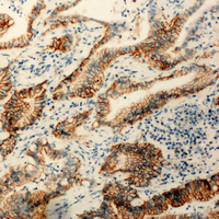 Immunohistochemical analysis of Claudin 3 staining in human colon cancer formalin fixed paraffin embedded tissue section. The section was pre-treated using heat mediated antigen retrieval with sodium citrate buffer (pH 6.0). The section was then incubated with the antibody at room temperature and detected using an HRP conjugated compact polymer system. DAB was used as the chromogen. The section was then counterstained with haematoxylin and mounted with DPX.
 -
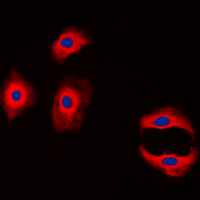 Immunofluorescent analysis of Claudin 3 staining in MCF7 cells. Formalin-fixed cells were permeabilized with 0.1% Triton X-100 in TBS for 5-10 minutes and blocked with 3% BSA-PBS for 30 minutes at room temperature. Cells were probed with the primary antibody in 3% BSA-PBS and incubated overnight at 4 °C in a hidified chamber. Cells were washed with PBST and incubated with a DyLight 594-conjugated secondary antibody (red) in PBS at room temperature in the dark. DAPI was used to stain the cell nuclei (blue).
An Informatics-assisted Label-free Approach for Personalized Tissue Membrane Proteomics: Case Study on Colorectal Cancer
CD24 regulated gene expression and distribution of tight junction proteins is associated with altered barrier function in oral epithelial monolayers
Inorganic arsenic exposure promotes malignant progression by HDAC6‐mediated down‐regulation of HTRA1