
 Anti-CDK5 (Phospho-Y15) Antibody
  Datasheet
 MSDS
MSDS
Description: Rabbit polyclonal antibody to CDK5 (Phospho-Y15) Immunogen: KLH-conjugated synthetic phosphopeptide corresponding to residues surrounding Y15 of human CDK5 protein. The exact sequence is proprietary. Purification: The antibody was purified by immunogen affinity chromatography. Clonality: Polyclonal Form: Liquid in 0.42% Potassium phosphate, 0.87% Sodium chloride, pH 7.3, 30% glycerol, and 0.01% sodium azide. Dilution: WB (1/500 - 1/1000), IH (1/100 - 1/200), IF/IC (1/100 - 1/500) Gene Symbol: CDK5 Alternative Names: CDKN5; Cyclin-dependent kinase 5; Cell division protein kinase 5; Serine/threonine-protein kinase PSSALRE; Tau protein kinase II catalytic subunit; TPKII catalytic subunitEntrez Gene (Human): 1020Entrez Gene (Mouse) : 12568Entrez Gene (Rat) : 140908SwissProt (Human): Q00535SwissProt (Mouse) : P49615SwissProt (Rat) : Q03114Storage/Stability : Shipped at 4°C. Upon delivery aliquot and store at -20°C for one year. Avoid freeze/thaw cycles.
-
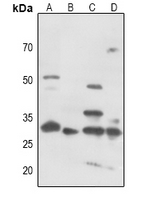 Western blot analysis of CDK5 (Phospho-Y15) expression in HEK293T (A), mouse muscle (B), mouse lung (C), rat muscle (D) whole cell lysates.
 -
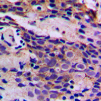 Immunohistochemical analysis of CDK5 (Phospho-Y15) staining in human breast cancer formalin fixed paraffin embedded tissue section. The section was pre-treated using heat mediated antigen retrieval with sodium citrate buffer (pH 6.0). The section was then incubated with the antibody at room temperature and detected using an HRP conjugated compact polymer system. DAB was used as the chromogen. The section was then counterstained with haematoxylin and mounted with DPX.
 -
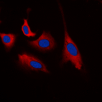 Immunofluorescent analysis of CDK5 (Phospho-Y15) staining in HEK293T cells. Formalin-fixed cells were permeabilized with 0.1% Triton X-100 in TBS for 5-10 minutes and blocked with 3% BSA-PBS for 30 minutes at room temperature. Cells were probed with the primary antibody in 3% BSA-PBS and incubated overnight at 4 °C in a hidified chamber. Cells were washed with PBST and incubated with a DyLight 594-conjugated secondary antibody (red) in PBS at room temperature in the dark. DAPI was used to stain the cell nuclei (blue).
 -
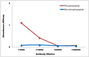 Direct ELISA antibody dose-response curve using Anti-CDK5 (Phospho-Y15) Antibody. Antigen (Phosphopeptide and non-phosphopeptide) concentration is 5 ug/ml. Goat Anti-Rabbit IgG (H&L) - HRP was used as the secondary antibody, and signal was developed by TMB substrate.
Phosphorylation of cyclin-dependent kinase 5 (Cdk5) at Tyr-15 is inhibited by Cdk5 activators and does not contribute to the activation of Cdk5
EGFR/MEK/ERK/CDK5-dependent integrin-independent FAK phosphorylated on serine 732 contributes to microtubule depolymerization and mitosis in tumor cells
Valproate reduces tau phosphorylation via cyclin-dependent kinase 5 and glycogen synthase kinase 3 signaling pathways