
 Anti-BCLX (Phospho-S62) Antibody
  Datasheet
 MSDS
MSDS
Description: Rabbit polyclonal antibody to BCLX (Phospho-S62) Immunogen: KLH-conjugated synthetic phosphopeptide corresponding to residues surrounding S62 of human BCLX protein. The exact sequence is proprietary. Purification: The antibody was purified by immunogen affinity chromatography. Clonality: Polyclonal Form: Liquid in 0.42% Potassium phosphate, 0.87% Sodium chloride, pH 7.3, 30% glycerol, and 0.01% sodium azide. Dilution: WB (1/500 - 1/1000), IH (1/100 - 1/200), IF/IC (1/100 - 1/500) Gene Symbol: BCL2L1 Alternative Names: BCL2L; BCLX; Bcl-2-like protein 1; Bcl2-L-1; Apoptosis regulator Bcl-XEntrez Gene (Human): 598Entrez Gene (Mouse) : 12048Entrez Gene (Rat) : 24888SwissProt (Human): Q07817SwissProt (Mouse) : Q64373SwissProt (Rat) : P53563Storage/Stability : Shipped at 4°C. Upon delivery aliquot and store at -20°C for one year. Avoid freeze/thaw cycles.
-
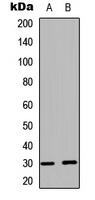 Western blot analysis of BCLX (Phospho-S62) expression in HEK293T UV-treated (A), rat muscle (B) whole cell lysates.
 -
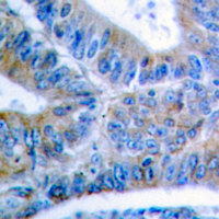 Immunohistochemical analysis of BCLX (Phospho-S62) staining in human colon cancer formalin fixed paraffin embedded tissue section. The section was pre-treated using heat mediated antigen retrieval with sodium citrate buffer (pH 6.0). The section was then incubated with the antibody at room temperature and detected using an HRP conjugated compact polymer system. DAB was used as the chromogen. The section was then counterstained with haematoxylin and mounted with DPX.
 -
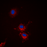 Immunofluorescent analysis of BCLX (Phospho-S62) staining in HEK293T cells. Formalin-fixed cells were permeabilized with 0.1% Triton X-100 in TBS for 5-10 minutes and blocked with 3% BSA-PBS for 30 minutes at room temperature. Cells were probed with the primary antibody in 3% BSA-PBS and incubated overnight at 4 °C in a hidified chamber. Cells were washed with PBST and incubated with a DyLight 594-conjugated secondary antibody (red) in PBS at room temperature in the dark. DAPI was used to stain the cell nuclei (blue).
 -
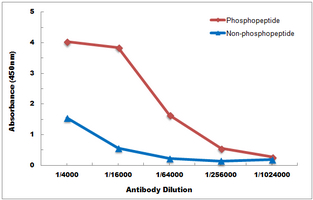 Direct ELISA antibody dose-response curve using Anti-BCLX (Phospho-S62) Antibody. Antigen (Phosphopeptide and non-phosphopeptide) concentration is 5 ug/ml. Goat Anti-Rabbit IgG (H&L) - HRP was used as the secondary antibody, and signal was developed by TMB substrate.
Critical role of anti-apoptotic Bcl-2 protein phosphorylation in mitotic death
ABT-737, a small molecule Bcl-2/Bcl-xL antagonist, increases antimitotic-mediated apoptosis in human prostate cancer cells
Cyclin-dependent Kinase-1 (Cdk1)/Cyclin B1 Dictates Cell Fate after Mitotic Arrest via Phosphoregulation of Antiapoptotic Bcl-2 Proteins
Tyrosine-phosphorylated caveolin-1 (Tyr-14) increases sensitivity to paclitaxel by inhibiting BCL2 and BCLxL proteins via c-Jun N-terminal kinase (JNK)