
 Anti-Lamin A/C (Phospho-S392) Antibody
  Datasheet
 MSDS
MSDS
Description: Rabbit polyclonal antibody to Lamin A/C (Phospho-S392) Immunogen: KLH-conjugated synthetic phosphopeptide corresponding to residues surrounding S392 of human Lamin A/C protein. The exact sequence is proprietary. Purification: The antibody was purified by immunogen affinity chromatography. Clonality: Polyclonal Form: Liquid in 0.42% Potassium phosphate, 0.87% Sodium chloride, pH 7.3, 30% glycerol, and 0.01% sodium azide. Dilution: WB (1/500 - 1/1000), IH (1/50 - 1/100), IF/IC (1/50 - 1/200) Gene Symbol: LMNA Alternative Names: LMN1; Prelamin-A/CEntrez Gene (Human): 4000Entrez Gene (Mouse) : 16905Entrez Gene (Rat) : 60374SwissProt (Human): P02545SwissProt (Mouse) : P48678SwissProt (Rat) : P48679Storage/Stability : Shipped at 4°C. Upon delivery aliquot and store at -20°C for one year. Avoid freeze/thaw cycles.
-
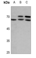 Western blot analysis of Lamin A/C (Phospho-S392) expression in DLD (A), mouse kidney (B), rat lung (C) whole cell lysates.
 -
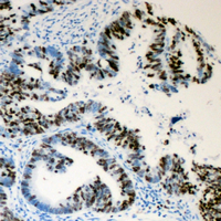 Immunohistochemical analysis of Lamin A/C (Phospho-S392) staining in human colon cancer formalin fixed paraffin embedded tissue section. The section was pre-treated using heat mediated antigen retrieval with sodium citrate buffer (pH 6.0). The section was then incubated with the antibody at room temperature and detected using an HRP conjugated compact polymer system. DAB was used as the chromogen. The section was then counterstained with haematoxylin and mounted with DPX.
 -
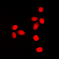 Immunofluorescent analysis of Lamin A/C (Phospho-S392) staining in SHSY5Y cells. Formalin-fixed cells were permeabilized with 0.1% Triton X-100 in TBS for 5-10 minutes and blocked with 3% BSA-PBS for 30 minutes at room temperature. Cells were probed with the primary antibody in 3% BSA-PBS and incubated overnight at 4 °C in a humidified chamber. Cells were washed with PBST and incubated with a DyLight 594-conjugated secondary antibody (red) in PBS at room temperature in the dark.
Cdk1, but not Cdk2, is the sole Cdk that is essential and sufficient to drive resumption of meiosis in mouse oocytes