
 Anti-HIF1 alpha Antibody
  Datasheet
 MSDS
MSDS
Description: Rabbit polyclonal antibody to HIF1 alpha Immunogen: KLH-conjugated synthetic peptide encompassing a sequence within the center region of human HIF1 alpha. The exact sequence is proprietary. Purification: The antibody was purified by immunogen affinity chromatography. Clonality: Polyclonal Form: Liquid in 0.42% Potassium phosphate, 0.87% Sodium chloride, pH 7.3, 30% glycerol, and 0.01% sodium azide. Dilution: WB (1/500 - 1/1000), IH (1/50 - 1/100), IF/IC (1/50 - 1/200) Gene Symbol: HIF1A Alternative Names: BHLHE78; MOP1; PASD8; Hypoxia-inducible factor 1-alpha; HIF-1-alpha; HIF1-alpha; ARNT-interacting protein; Basic-helix-loop-helix-PAS protein MOP1; Class E basic helix-loop-helix protein 78; bHLHe78; Member of PAS protein 1; PAS domain-containing protein 8Entrez Gene (Human): 3091Entrez Gene (Mouse) : 15251Entrez Gene (Rat) : 29560SwissProt (Human): Q16665SwissProt (Mouse) : Q61221SwissProt (Rat) : O35800Storage/Stability : Shipped at 4°C. Upon delivery aliquot and store at -20°C for one year. Avoid freeze/thaw cycles.
-
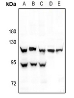 Western blot analysis of HIF1 alpha expression in HEK293T (A), A549 (B), MCF7 (C), H9C2 (D), SP20 (E) whole cell lysates.
 -
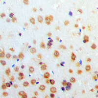 Immunohistochemical analysis of HIF1 alpha staining in human brain formalin fixed paraffin embedded tissue section. The section was pre-treated using heat mediated antigen retrieval with sodium citrate buffer (pH 6.0). The section was then incubated with the antibody at room temperature and detected using an HRP conjugated compact polymer system. DAB was used as the chromogen. The section was then counterstained with haematoxylin and mounted with DPX.
 -
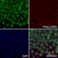 Immunofluorescent analysis of HIF1 alpha staining in LO2 cells. Formalin-fixed cells were permeabilized with 0.1% Triton X-100 in TBS for 5-10 minutes and blocked with 3% BSA-PBS for 30 minutes at room temperature. Cells were probed with the primary antibody in 3% BSA-PBS and incubated overnight at 4 °C in a hidified chamber. Cells were washed with PBST and incubated with a AF488-conjugated secondary antibody (green) in PBS at room temperature in the dark. Phalloidin - AF594 was used to stain Actin filaments (red). DAPI was used to stain the cell nuclei (blue).
Effect of αvβ3 Blockade Against Acute Lung Injury Induced by Influenza A Virus and Its Mechanism
Zi-Su-Zi decoction improves airway hyperresponsiveness in cough-variant asthma rat model through PI3K/AKT1/mTOR, JAK2/STAT3 and HIF-1α/NF-κB signaling pathways