
 Anti-Renin Receptor Antibody
  Datasheet
 MSDS
MSDS
Description: Rabbit polyclonal antibody to Renin Receptor Immunogen: KLH-conjugated synthetic peptide encompassing a sequence within the center region of human Renin Receptor. The exact sequence is proprietary. Purification: The antibody was purified by immunogen affinity chromatography. Clonality: Polyclonal Form: Liquid in 0.42% Potassium phosphate, 0.87% Sodium chloride, pH 7.3, 30% glycerol, and 0.01% sodium azide. Dilution: WB (1/500 - 1/1000), IH (1/50 - 1/100), IF/IC (1/50 - 1/200) Gene Symbol: ATP6AP2 Alternative Names: ATP6IP2; CAPER; ELDF10; Renin receptor; ATPase H(+)-transporting lysosomal accessory protein 2; ATPase H(+)-transporting lysosomal-interacting protein 2; ER-localized type I transmembrane adaptor; Embryonic liver differentiation factor 10; N14F; Renin/prorenin receptor; Vacuolar ATP synthase membrane sector-associated protein M8-9; ATP6M8-9; V-ATPase M8.9 subunitEntrez Gene (Human): 10159Entrez Gene (Mouse) : 70495Entrez Gene (Rat) : 302526SwissProt (Human): O75787SwissProt (Mouse) : Q9CYN9SwissProt (Rat) : Q6AXS4Storage/Stability : Shipped at 4°C. Upon delivery aliquot and store at -20°C for one year. Avoid freeze/thaw cycles.
-
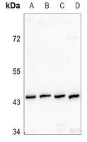 Western blot analysis of Renin Receptor expression in THP1 (A), HEK293T (B), Hela (C), CT26 (D) whole cell lysates.
 -
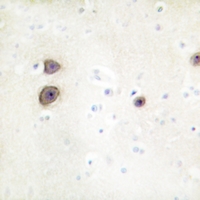 Immunohistochemical analysis of Renin Receptor staining in human brain formalin fixed paraffin embedded tissue section. The section was pre-treated using heat mediated antigen retrieval with sodium citrate buffer (pH 6.0). The section was then incubated with the antibody at room temperature and detected using an HRP conjugated compact polymer system. DAB was used as the chromogen. The section was then counterstained with haematoxylin and mounted with DPX.
 -
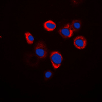 Immunofluorescent analysis of Renin Receptor staining in Raw264.7 cells. Formalin-fixed cells were permeabilized with 0.1% Triton X-100 in TBS for 5-10 minutes and blocked with 3% BSA-PBS for 30 minutes at room temperature. Cells were probed with the primary antibody in 3% BSA-PBS and incubated overnight at 4 °C in a humidified chamber. Cells were washed with PBST and incubated with a DyLight 594-conjugated secondary antibody (red) in PBS at room temperature in the dark. DAPI was used to stain the cell nuclei (blue).
Nuclear expression of renin-angiotensin system components in NRK-52E renal epithelial cells