
 Anti-p63 Antibody
  Datasheet
 MSDS
MSDS
Description: Rabbit polyclonal antibody to p63 Immunogen: KLH-conjugated synthetic peptide encompassing a sequence within the C-term region of human p63. The exact sequence is proprietary. Purification: The antibody was purified by immunogen affinity chromatography. Clonality: Polyclonal Form: Liquid in 0.42% Potassium phosphate, 0.87% Sodium chloride, pH 7.3, 30% glycerol, and 0.01% sodium azide. Dilution: WB (1/500 - 1/1000), IH (1/100 - 1/200), IF/IC (1/100 - 1/500) Gene Symbol: TP63 Alternative Names: KET; P63; P73H; P73L; TP73L; Tumor protein 63; p63; Chronic ulcerative stomatitis protein; CUSP; Keratinocyte transcription factor KET; Transformation-related protein 63; TP63; Tumor protein p73-like; p73L; p40; p51Entrez Gene (Human): 8626Entrez Gene (Mouse) : 22061Entrez Gene (Rat) : 246334SwissProt (Human): Q9H3D4SwissProt (Mouse) : O88898SwissProt (Rat) : Q9JJP6Storage/Stability : Shipped at 4°C. Upon delivery aliquot and store at -20°C for one year. Avoid freeze/thaw cycles.
-
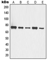 Western blot analysis of p63 expression in A549 (A), HCT116 (B), COLO205 (C), LOVO (D), HT29 (E) whole cell lysates.
 -
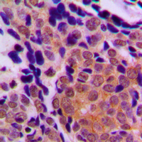 Immunohistochemical analysis of p63 staining in human breast cancer formalin fixed paraffin embedded tissue section. The section was pre-treated using heat mediated antigen retrieval with sodium citrate buffer (pH 6.0). The section was then incubated with the antibody at room temperature and detected using an HRP conjugated compact polymer system. DAB was used as the chromogen. The section was then counterstained with haematoxylin and mounted with DPX.
 -
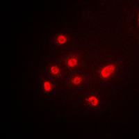 Immunofluorescent analysis of p63 staining in A549 cells. Formalin-fixed cells were permeabilized with 0.1% Triton X-100 in TBS for 5-10 minutes and blocked with 3% BSA-PBS for 30 minutes at room temperature. Cells were probed with the primary antibody in 3% BSA-PBS and incubated overnight at 4 °C in a humidified chamber. Cells were washed with PBST and incubated with a DyLight 594-conjugated secondary antibody (red) in PBS at room temperature in the dark.
Sp6 and Sp8 Transcription Factors Control AER Formation and Dorsal-Ventral Patterning in Limb Development
Adipose-derived stem cell to epithelial stem cell transdifferentiation: a mechanism to potentially improve understanding of fat grafting's impact on skin rejuvenation
Transcriptional profiling of epidermal barrier formation in vitro
p63 attenuates epithelial to mesenchymal potential in an experimental prostate cell model
Interaction of the oncogenic miR-21 microRNA and the p53 tumor suppressor pathway
Altered REDD1, myostatin, and Akt/mTOR/FoxO/MAPK signaling in streptozotocin-induced diabetic muscle atrophy
Morphogenesis of the developing mammary gland: stage-dependent impact of adipocytes
CD44posCD49fhiCD133/2hi defines xenograft-initiating cells in estrogen receptor-negative breast cancer