
 Anti-p53 (pS15) Antibody
  Datasheet
 MSDS
MSDS
Description: Rabbit polyclonal antibody to p53 (Phospho-S15) Immunogen: KLH-conjugated synthetic phosphopeptide corresponding to residues surrounding S15 of human p53 protein. The exact sequence is proprietary. Purification: The antibody was purified by immunogen affinity chromatography. Clonality: Polyclonal Form: Liquid in 0.42% Potassium phosphate, 0.87% Sodium chloride, pH 7.3, 30% glycerol, and 0.01% sodium azide. Dilution: WB (1/500 - 1/1000), IH (1/100 - 1/200), IF/IC (1/100 - 1/500), IP (1/10 - 1/100) Gene Symbol: TP53 Alternative Names: P53; Cellular tumor antigen p53; Antigen NY-CO-13; Phosphoprotein p53; Tumor suppressor p53Entrez Gene (Human): 7157Entrez Gene (Rat) : 24842SwissProt (Human): P04637SwissProt (Rat) : P10361Storage/Stability : Shipped at 4°C. Upon delivery aliquot and store at -20°C for one year. Avoid freeze/thaw cycles.
-
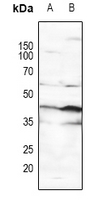 Western blot analysis of p53 (Phospho-S15) expression in Hela (A), Jurkat (B) whole cell lysates.
 -
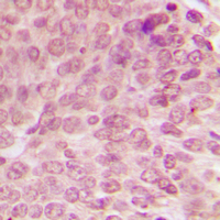 Immunohistochemical analysis of p53 (Phospho-S15) staining in human breast cancer formalin fixed paraffin embedded tissue section. The section was pre-treated using heat mediated antigen retrieval with sodium citrate buffer (pH 6.0). The section was then incubated with the antibody at room temperature and detected using an HRP conjugated compact polymer system. DAB was used as the chromogen. The section was then counterstained with haematoxylin and mounted with DPX.
 -
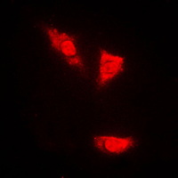 Immunofluorescent analysis of p53 (Phospho-S15) staining in PC12 cells. Formalin-fixed cells were permeabilized with 0.1% Triton X-100 in TBS for 5-10 minutes and blocked with 3% BSA-PBS for 30 minutes at room temperature. Cells were probed with the primary antibody in 3% BSA-PBS and incubated overnight at 4 °C in a humidified chamber. Cells were washed with PBST and incubated with a DyLight 594-conjugated secondary antibody (red) in PBS at room temperature in the dark.
 -
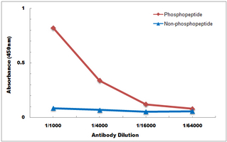 Direct ELISA antibody dose-response curve using Anti-p53 (Phospho-S15) Antibody. Antigen (Phosphopeptide and non-phosphopeptide) concentration is 5 ug/ml. Goat Anti-Rabbit IgG (H&L) - HRP was used as the secondary antibody, and signal was developed by TMB substrate.
Chromatin structure, transcriptional activity and DNA repair efficiency affect the outcome of chemotherapy in multiple myeloma
SETD2 is required for DNA double-strand break repair and activation of the p53-mediated checkpoint
DIM (3,3'-diindolylmethane) confers protection against ionizing radiation by a unique mechanism
Progressive changes in chromatin structure and DNA damage response signals in bone marrow and peripheral blood during myelomagenesis