
 Anti-AP2 alpha/beta Antibody
  Datasheet
 MSDS
MSDS
Description: Rabbit polyclonal antibody to AP2 alpha/beta Immunogen: KLH-conjugated synthetic peptide encompassing a sequence within the C-term region of human AP2 alpha/beta. The exact sequence is proprietary. Purification: The antibody was purified by immunogen affinity chromatography. Clonality: Polyclonal Form: Liquid in 0.42% Potassium phosphate, 0.87% Sodium chloride, pH 7.3, 30% glycerol, and 0.01% sodium azide. Dilution: WB (1/500 - 1/1000), IH (1/100 - 1/200), IF/IC (1/100 - 1/500), ChIP (1/100 - 1/500), EMSA (Use at an assay dependent dilution) Gene Symbol: TFAP2A; TFAP2B Alternative Names: TFAP2A; AP2TF; TFAP2; Transcription factor AP-2-alpha; AP2-alpha; AP-2 transcription factor; Activating enhancer-binding protein 2-alpha; Activator protein 2; AP-2; TFAP2B; Transcription factor AP-2-beta; AP2-beta; Activating enhancer-binding protein 2-betaEntrez Gene (Human): 7020Entrez Gene (Mouse) : 21418; 21419SwissProt (Human): P05549; Q92481SwissProt (Mouse) : P34056; Q61313SwissProt (Rat) : P58197Storage/Stability : Shipped at 4°C. Upon delivery aliquot and store at -20°C for one year. Avoid freeze/thaw cycles.
-
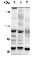 Western blot analysis of AP2 alpha/beta expression in PC3 (A), MCF7 (B), Myla2059 (C) whole cell lysates.
 -
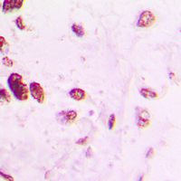 Immunohistochemical analysis of AP2 alpha/beta staining in human lung cancer formalin fixed paraffin embedded tissue section. The section was pre-treated using heat mediated antigen retrieval with sodium citrate buffer (pH 6.0). The section was then incubated with the antibody at room temperature and detected using an HRP conjugated compact polymer system. DAB was used as the chromogen. The section was then counterstained with haematoxylin and mounted with DPX.
 -
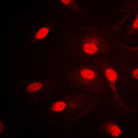 Immunofluorescent analysis of AP2 alpha/beta staining in HepG2 cells. Formalin-fixed cells were permeabilized with 0.1% Triton X-100 in TBS for 5-10 minutes and blocked with 3% BSA-PBS for 30 minutes at room temperature. Cells were probed with the primary antibody in 3% BSA-PBS and incubated overnight at 4 °C in a humidified chamber. Cells were washed with PBST and incubated with a DyLight 594-conjugated secondary antibody (red) in PBS at room temperature in the dark.
 -
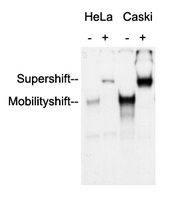 Anti-AP2 alpha/beta Antibody was used in an Electrophoretic Mobility Shift Assay (EMSA) to supershift the protein-DNA complex. Radiolabelled, double-stranded DNA oligonucleotides (10.000 cpm per lane) harbouring a binding site for AP2 alpha/beta were incubated with each 2 ug of nuclear extract (NE) from HeLa and Caski cells, respectively. Samples were incubated for 30 minutes at room temperature to allow the formation of protein-DNA complexes. Anti-AP2 alpha/beta Antibody were added to the samples (as indicated) and incubated for further 60 minutes at 4°C. Samples were separated in a 5.5% PAGE. The Gel was dried under vacuum and for autoradiography a X-ray film was exposed with an intensifying screen for 2 days at -80°C. Specific protein-DNA complexes were quantitatively supershifted with Anti-AP2 alpha/beta Antibody, verifying the binding of AP2 alpha/beta to the DNA oligonucleotide.
 -
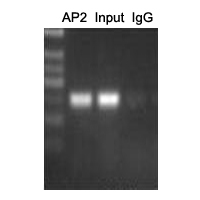 ChIP analysis of Cervical cancer cell lines lysate, incubated for 12 hours at 4°C. Cross-linking (X-ChIP) using formaldehyde for 10 minutes. Detection step: Semiquantitative PCR. Positive control: Tumor cell lines Hela. Negative control: Human primary keratinocytes.
ID1 promotes breast cancer metastasis by S100A9 regulation
A polymorphism in IRF4 affects human pigmentation through a tyrosinase-dependent MITF/TFAP2A pathway
SOX10 directly modulates ERBB3 transcription via an intronic neural crest enhancer
Transcriptional regulation of the human p450 oxidoreductase gene: hormonal regulation and influence of promoter polymorphisms
Transcriptional inhibition of intestinal NHE8 expression by glucocorticoids involves Pax5
Journal
AM J PHYSIOL-GASTR L
IF
4.5
Application
Gel supershift assays
Reactivity
Human
PMID
20671194
Upregulating CD59: a new strategy for protection of neurons from complement-mediated degeneration