
 Anti-NF-kappaB p65 (Phospho-S536) Antibody
  Datasheet
 MSDS
MSDS
Description: Rabbit polyclonal antibody to NF-kappaB p65 (Phospho-S536) Immunogen: KLH-conjugated synthetic phosphopeptide corresponding to residues surrounding S536 of human NF-kappaB p65 protein. The exact sequence is proprietary. Purification: The antibody was purified by immunogen affinity chromatography. Clonality: Polyclonal Form: Liquid in 0.42% Potassium phosphate, 0.87% Sodium chloride, pH 7.3, 30% glycerol, and 0.01% sodium azide. Dilution: WB (1/500 - 1/1000), IH (1/50 - 1/200), IF/IC (1/50 - 1/200), IP (1/10 - 1/100) Gene Symbol: RELA Alternative Names: NFKB3; Transcription factor p65; Nuclear factor NF-kappa-B p65 subunit; Nuclear factor of kappa light polypeptide gene enhancer in B-cells 3Entrez Gene (Human): 5970Entrez Gene (Mouse) : 19697SwissProt (Human): Q04206SwissProt (Mouse) : Q04207Storage/Stability : Shipped at 4°C. Upon delivery aliquot and store at -20°C for one year. Avoid freeze/thaw cycles.
-
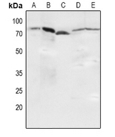 Western blot analysis of NF-kappaB p65 (Phospho-S536) expression in Hela (A), HEK293T (B), NIH3T3 (C), mouse kidney (D), H9C2 (E) whole cell lysates.
 -
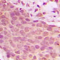 Immunohistochemical analysis of NF-kappaB p65 (Phospho-S536) staining in human breast cancer formalin fixed paraffin embedded tissue section. The section was pre-treated using heat mediated antigen retrieval with sodium citrate buffer (pH 6.0). The section was then incubated with the antibody at room temperature and detected using an HRP conjugated compact polymer system. DAB was used as the chromogen. The section was then counterstained with haematoxylin and mounted with DPX.
 -
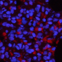 Immunofluorescent analysis of NF-kappaB p65 (Phospho-S536) staining in rat lung. The section was pre-treated using heat mediated antigen retrieval with sodium citrate buffer (pH 6.0). The section was then incubated with the antibody at room temperature and detected using a AF594-conjugated secondary antibody (red) in PBS at room temperature in the dark. DAPI was used to stain the cell nuclei (blue).
 -
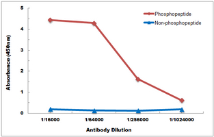 Direct ELISA antibody dose-response curve using Anti-NF-kappaB p65 (Phospho-S536) Antibody. Antigen (Phosphopeptide and non-phosphopeptide) concentration is 5 ug/ml. Goat Anti-Rabbit IgG (H&L) - HRP was used as the secondary antibody, and signal was developed by TMB substrate.
NLRP3 Gene Silencing Ameliorates Diabetic Cardiomyopathy in a Type 2 Diabetes Rat Model
NF-?B p65 phosphorylated at serine-536 is an independent prognostic factor in Swedish colorectal cancer patients
CD40 Is Essential in the Upregulation of TRAF Proteins and NF-KappaB-Dependent Proinflammatory Gene Expression after Arterial Injury
Zi-Su-Zi decoction improves airway hyperresponsiveness in cough-variant asthma rat model through PI3K/AKT1/mTOR, JAK2/STAT3 and HIF-1α/NF-κB signaling pathways