
 Anti-PDGFR beta Antibody
  Datasheet
 MSDS
MSDS
Description: Rabbit polyclonal antibody to PDGFR beta Immunogen: KLH-conjugated synthetic peptide encompassing a sequence within the center region of human PDGFR beta. The exact sequence is proprietary. Purification: The antibody was purified by immunogen affinity chromatography. Clonality: Polyclonal Form: Liquid in 0.42% Potassium phosphate, 0.87% Sodium chloride, pH 7.3, 30% glycerol, and 0.01% sodium azide. Dilution: WB (1/500 - 1/1000), IH (1/500 - 1/200), IF/IC (1/50 - 1/200) Gene Symbol: PDGFRB Alternative Names: PDGFR; PDGFR1; Platelet-derived growth factor receptor beta; PDGF-R-beta; PDGFR-beta; Beta platelet-derived growth factor receptor; Beta-type platelet-derived growth factor receptor; CD140 antigen-like family member B; Platelet-derived growth factor receptor 1; PDGFR-1; CD140bEntrez Gene (Human): 5159Entrez Gene (Mouse) : 18596Entrez Gene (Rat) : 24629SwissProt (Human): P09619SwissProt (Mouse) : P05622SwissProt (Rat) : Q05030Storage/Stability : Shipped at 4°C. Upon delivery aliquot and store at -20°C for one year. Avoid freeze/thaw cycles.
-
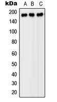 Western blot analysis of PDGFR beta expression in SP2/0 (A), H9C2 (B), PC12 (C) whole cell lysates.
 -
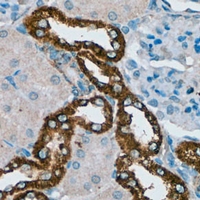 Immunohistochemical analysis of PDGFR beta staining in rat kidney formalin fixed paraffin embedded tissue section. The section was pre-treated using heat mediated antigen retrieval with sodium citrate buffer (pH 6.0). The section was then incubated with the antibody at room temperature and detected using an HRP conjugated compact polymer system. DAB was used as the chromogen. The section was then counterstained with haematoxylin and mounted with DPX.
 -
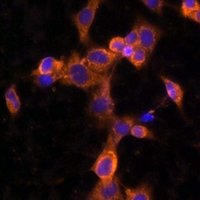 Immunofluorescent analysis of PDGFR beta staining in NIH3T3 cells. Formalin-fixed cells were permeabilized with 0.1% Triton X-100 in TBS for 5-10 minutes and blocked with 3% BSA-PBS for 30 minutes at room temperature. Cells were probed with the primary antibody in 3% BSA-PBS and incubated overnight at 4 °C in a humidified chamber. Cells were washed with PBST and incubated with a AF594-conjugated secondary antibody (red) in PBS at room temperature in the dark. DAPI was used to stain the cell nuclei (blue).
Bone marrow multipotent mesenchymal stroma cells act as pericyte-like migratory vehicles in experimental gliomas
A feed-forward mechanosignaling loop confers resistance to therapies targeting the MAPK pathway in BRAF-mutant melanoma
Protocatechuic aldehyde increases pericyte coverage and mitigates pericyte damage to enhance the atherosclerotic plaque stability