
 Anti-LCK (Phospho-Y393) Antibody
  Datasheet
 MSDS
MSDS
Description: Rabbit polyclonal antibody to LCK (Phospho-Y393) Immunogen: KLH-conjugated synthetic phosphopeptide corresponding to residues surrounding Y393 of human LCK protein. The exact sequence is proprietary. Purification: The antibody was purified by immunogen affinity chromatography. Clonality: Polyclonal Form: Liquid in 0.42% Potassium phosphate, 0.87% Sodium chloride, pH 7.3, 30% glycerol, and 0.01% sodium azide. Dilution: WB (1/500 - 1/1000), IF/IC (1/100 - 1/500) Gene Symbol: LCK Alternative Names: Tyrosine-protein kinase Lck; Leukocyte C-terminal Src kinase; LSK; Lymphocyte cell-specific protein-tyrosine kinase; Protein YT16; Proto-oncogene Lck; T cell-specific protein-tyrosine kinase; p56-LCKEntrez Gene (Human): 3932Entrez Gene (Mouse) : 16818Entrez Gene (Rat) : 313050SwissProt (Human): P06239SwissProt (Mouse) : P06240SwissProt (Rat) : Q01621Storage/Stability : Shipped at 4°C. Upon delivery aliquot and store at -20°C for one year. Avoid freeze/thaw cycles.
-
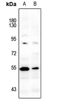 Western blot analysis of LCK (Phospho-Y393) expression in Myla2059 (A), HuT78 (B) whole cell lysates.
 -
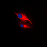 Immunofluorescent analysis of LCK (Phospho-Y393) staining in MCF7 cells. Formalin-fixed cells were permeabilized with 0.1% Triton X-100 in TBS for 5-10 minutes and blocked with 3% BSA-PBS for 30 minutes at room temperature. Cells were probed with the primary antibody in 3% BSA-PBS and incubated overnight at 4 °C in a humidified chamber. Cells were washed with PBST and incubated with a DyLight 594-conjugated secondary antibody (red) in PBS at room temperature in the dark. DAPI was used to stain the cell nuclei (blue).
 -
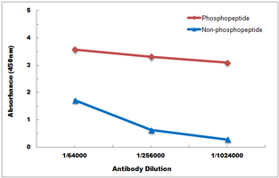 Direct ELISA antibody dose-response curve using Anti-LCK (Phospho-Y393) Antibody. Antigen (Phosphopeptide and non-phosphopeptide) concentration is 5 ug/ml. Goat Anti-Rabbit IgG (H&L) - HRP was used as the secondary antibody, and signal was developed by TMB substrate.