
 Anti-Cytokeratin 5 Antibody
  Datasheet
 MSDS
MSDS
Description: Rabbit polyclonal antibody to Cytokeratin 5 Immunogen: KLH-conjugated synthetic peptide encompassing a sequence within the C-term region of human Cytokeratin 5. The exact sequence is proprietary. Purification: The antibody was purified by immunogen affinity chromatography. Clonality: Polyclonal Form: Liquid in 0.42% Potassium phosphate, 0.87% Sodium chloride, pH 7.3, 30% glycerol, and 0.01% sodium azide. Dilution: WB (1/500 - 1/1000), IH (1/50 - 1/100), IF/IC (1/50 - 1/200), FC (1/100 - 1/200) Gene Symbol: KRT5 Alternative Names: Keratin, type II cytoskeletal 5; 58 kDa cytokeratin; Cytokeratin-5; CK-5; Keratin-5; K5; Type-II keratin Kb5Entrez Gene (Human): 3852Entrez Gene (Mouse) : 110308Entrez Gene (Rat) : 369017SwissProt (Human): P13647SwissProt (Mouse) : Q922U2SwissProt (Rat) : Q6P6Q2Storage/Stability : Shipped at 4°C. Upon delivery aliquot and store at -20°C for one year. Avoid freeze/thaw cycles.
-
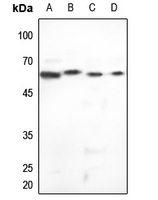 Western blot analysis of Cytokeratin 5 expression in HEK293T (A), Hela (B), mouse brain (C), rat brain (D) whole cell lysates.
 -
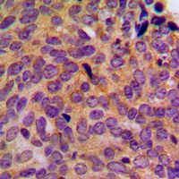 Immunohistochemical analysis of Cytokeratin 5 staining in human breast cancer formalin fixed paraffin embedded tissue section. The section was pre-treated using heat mediated antigen retrieval with sodium citrate buffer (pH 6.0). The section was then incubated with the antibody at room temperature and detected using an HRP conjugated compact polymer system. DAB was used as the chromogen. The section was then counterstained with haematoxylin and mounted with DPX.
 -
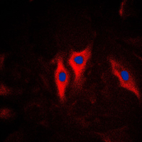 Immunofluorescent analysis of Cytokeratin 5 staining in HeLa cells. Formalin-fixed cells were permeabilized with 0.1% Triton X-100 in TBS for 5-10 minutes and blocked with 3% BSA-PBS for 30 minutes at room temperature. Cells were probed with the primary antibody in 3% BSA-PBS and incubated overnight at 4 °C in a humidified chamber. Cells were washed with PBST and incubated with a DyLight 594-conjugated secondary antibody (red) in PBS at room temperature in the dark. DAPI was used to stain the cell nuclei (blue).
PSGR promotes prostatic intraepithelial neoplasia and prostate cancer xenograft growth through NF-kB
Loss of primary cilia occurs early in breast cancer development
Epithelial basal cells are distinct from dendritic cells and macrophages in the mouse epididymis
Cessation of epithelial Bmp signaling switches the differentiation of crown epithelia to the root lineage in a ß-catenin-dependent manner
p63 attenuates epithelial to mesenchymal potential in an experimental prostate cell model
Estrogenic modulation of uropathogenic Escherichia coli infection pathogenesis in a murine menopause model
Evidence for a partial epithelial-mesenchymal transition in postnatal stages of rat auditory organ morphogenesis
Loss of transforming growth factor beta type II receptor increases aggressive tumor behavior and reduces survival in lung adenocarcinoma and squamous cell carcinoma
Conditional activation of Pik3ca(H1047R) in a knock-in mouse model promotes mammary tumorigenesis and emergence of mutations
Three differentiation states risk-stratify bladder cancer into distinct subtypes
Stromal cell networks regulate thymocyte migration and dendritic cell behavior in the thymus