
 Anti-Gamma-enolase Antibody
  Datasheet
 MSDS
MSDS
Description: Rabbit polyclonal antibody to Gamma-enolase Immunogen: KLH-conjugated synthetic peptide encompassing a sequence within the C-term region of human Gamma-enolase. The exact sequence is proprietary. Purification: The antibody was purified by immunogen affinity chromatography. Clonality: Polyclonal Form: Liquid in 0.42% Potassium phosphate, 0.87% Sodium chloride, pH 7.3, 30% glycerol, and 0.01% sodium azide. Dilution: WB (1/500 - 1/1000), IH (1/100 - 1/200), IF/IC (1/100 - 1/500), FC (1/100 - 1/200) Gene Symbol: ENO2 Alternative Names: Gamma-enolase; 2-phospho-D-glycerate hydro-lyase; Enolase 2; Neural enolase; Neuron-specific enolase; NSEEntrez Gene (Human): 2026Entrez Gene (Mouse) : 13807Entrez Gene (Rat) : 100911625; 24334SwissProt (Human): P09104SwissProt (Mouse) : P17183SwissProt (Rat) : P07323Storage/Stability : Shipped at 4°C. Upon delivery aliquot and store at -20°C for one year. Avoid freeze/thaw cycles.
-
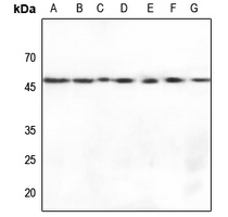 Western blot analysis of Gamma-enolase expression in HEK293T (A), Hela (B), U2OS (C), mouse brain (D), mouse eyes (E), rat brain (F), rat liver (G) whole cell lysates.
 -
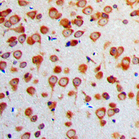 Immunohistochemical analysis of Gamma-enolase staining in human brain formalin fixed paraffin embedded tissue section. The section was pre-treated using heat mediated antigen retrieval with sodium citrate buffer (pH 6.0). The section was then incubated with the antibody at room temperature and detected using an HRP conjugated compact polymer system. DAB was used as the chromogen. The section was then counterstained with haematoxylin and mounted with DPX.
 -
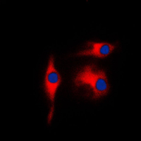 Immunofluorescent analysis of Gamma-enolase staining in K562 cells. Formalin-fixed cells were permeabilized with 0.1% Triton X-100 in TBS for 5-10 minutes and blocked with 3% BSA-PBS for 30 minutes at room temperature. Cells were probed with the primary antibody in 3% BSA-PBS and incubated overnight at 4 °C in a humidified chamber. Cells were washed with PBST and incubated with a DyLight 594-conjugated secondary antibody (red) in PBS at room temperature in the dark. DAPI was used to stain the cell nuclei (blue).
Deletion of KCC3 in parvalbumin neurons leads to locomotor deficit in a conditional mouse model of peripheral neuropathy associated with agenesis of the corpus callosum
The expression and significance of neuronal iconic proteins in podocytes
Potential of adipose-derived mesenchymal stem cells and skeletal muscle-derived satellite cells for somatic cell nuclear transfer mediated transgenesis in Arbas Cashmere goats
Acute minocycline treatment mitigates the symptoms of mild blast-induced traumatic brain injury
Stress and traumatic brain injury: a behavioral, proteomics, and histological study
Time-dependent changes in serum biomarker levels after blast traumatic brain injury
Siah2-Dependent Concerted Activity of HIF and FoxA2 Regulates Formation of Neuroendocrine Phenotype and Neuroendocrine Prostate Tumors
Transglutaminase 2 protects against ischemic stroke
Evaluation of blastomere biopsy using mouse model indicates the potential high-risk of neurodegenerative disorders in the offspring