
 Anti-E Cadherin Antibody
  Datasheet
 MSDS
MSDS
Description: Rabbit polyclonal antibody to E Cadherin Immunogen: KLH-conjugated synthetic peptide encompassing a sequence within the C-term region of human E Cadherin. The exact sequence is proprietary. Purification: The antibody was purified by immunogen affinity chromatography. Clonality: Polyclonal Form: Liquid in 0.42% Potassium phosphate, 0.87% Sodium chloride, pH 7.3, 30% glycerol, and 0.01% sodium azide. Dilution: WB (1/500 - 1/1000), IH (1/100 - 1/200), IF/IC (1/100 - 1/500), IP (1/10 - 1/100), FC (1/100 - 1/200) Gene Symbol: CDH1 Alternative Names: CDHE; UVO; Cadherin-1; CAM 120/80; Epithelial cadherin; E-cadherin; Uvomorulin; CD324Entrez Gene (Human): 999Entrez Gene (Mouse) : 12550Entrez Gene (Rat) : 83502SwissProt (Human): P12830SwissProt (Mouse) : P09803SwissProt (Rat) : Q9R0T4Storage/Stability : Shipped at 4°C. Upon delivery aliquot and store at -20°C for one year. Avoid freeze/thaw cycles.
-
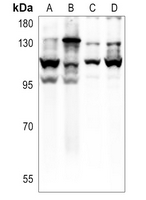 Western blot analysis of E Cadherin expression in A375 (A), HepG2 (B), mouse brain (C), rat brain (D) whole cell lysates.
 -
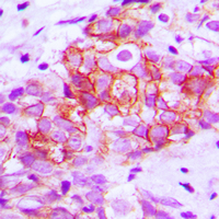 Immunohistochemical analysis of E Cadherin staining in human breast cancer formalin fixed paraffin embedded tissue section. The section was pre-treated using heat mediated antigen retrieval with sodium citrate buffer (pH 6.0). The section was then incubated with the antibody at room temperature and detected using an HRP conjugated compact polymer system. DAB was used as the chromogen. The section was then counterstained with haematoxylin and mounted with DPX.
 -
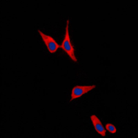 Immunofluorescent analysis of E Cadherin staining in PC12 cells. Formalin-fixed cells were permeabilized with 0.1% Triton X-100 in TBS for 5-10 minutes and blocked with 3% BSA-PBS for 30 minutes at room temperature. Cells were probed with the primary antibody in 3% BSA-PBS and incubated overnight at 4 °C in a humidified chamber. Cells were washed with PBST and incubated with a DyLight 594-conjugated secondary antibody (red) in PBS at room temperature in the dark. DAPI was used to stain the cell nuclei (blue).
 -
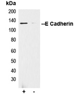 Immunoprecipitation of E Cadherin from 0.5mg HEK293F whole cell extract lysate, using 5ug of Anti-E Cadherin Antibody and 50ul of protein G magnetic beads (+). No antibody was added to the control (-). The antibody was incubated under agitation with Protein G beads for 10min, HEK293F whole cell extract lysate diluted in RIPA buffer was added to each sample and incubated for a further 10min under agitation. Proteins were eluted by addition of 40ul SDS loading buffer and incubated for 10min at 70°C; 10ul of each sample was separated on a SDS PAGE gel, transferred to a nitrocellulose membrane, blocked with 5% BSA and probed with Anti-E Cadherin Antibody.
Quantitative proteomics identifies myoferlin as a novel regulator of A Disintegrin and Metalloproteinase 12 in HeLa cells