
 Anti-Calnexin Antibody
  Datasheet
 MSDS
MSDS
Description: Rabbit polyclonal antibody to Calnexin Immunogen: KLH-conjugated synthetic peptide encompassing a sequence within the C-term region of human Calnexin. The exact sequence is proprietary. Purification: The antibody was purified by immunogen affinity chromatography. Clonality: Polyclonal Form: Liquid in 0.42% Potassium phosphate, 0.87% Sodium chloride, pH 7.3, 30% glycerol, and 0.01% sodium azide. Dilution: WB (1/500 - 1/1000), IH (1/50 - 1/100), IF/IC (1/50 - 1/200) Gene Symbol: CANX Alternative Names: Calnexin; IP90; Major histocompatibility complex class I antigen-binding protein p88; p90Entrez Gene (Human): 821Entrez Gene (Mouse) : 12330Entrez Gene (Rat) : 29144SwissProt (Human): P27824SwissProt (Mouse) : P35564SwissProt (Rat) : P35565Storage/Stability : Shipped at 4°C. Upon delivery aliquot and store at -20°C for one year. Avoid freeze/thaw cycles.
-
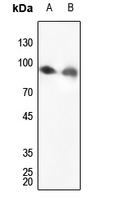 Western blot analysis of Calnexin expression in Hela (A), mouse lung (B) whole cell lysates.
 -
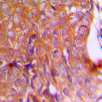 Immunohistochemical analysis of Calnexin staining in human breast cancer formalin fixed paraffin embedded tissue section. The section was pre-treated using heat mediated antigen retrieval with sodium citrate buffer (pH 6.0). The section was then incubated with the antibody at room temperature and detected using an HRP conjugated compact polymer system. DAB was used as the chromogen. The section was then counterstained with haematoxylin and mounted with DPX.
 -
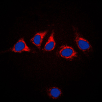 Immunofluorescent analysis of Calnexin staining in HeLa cells. Formalin-fixed cells were permeabilized with 0.1% Triton X-100 in TBS for 5-10 minutes and blocked with 3% BSA-PBS for 30 minutes at room temperature. Cells were probed with the primary antibody in 3% BSA-PBS and incubated overnight at 4 °C in a humidified chamber. Cells were washed with PBST and incubated with a DyLight 594-conjugated secondary antibody (red) in PBS at room temperature in the dark. DAPI was used to stain the cell nuclei (blue).
Quantitative proteomics and biochemical analyses reveal the role of endoplasmin in the regulation of the expression and secretion of A Disintegrin And Metalloproteinase 12
Mycobacterium tuberculosis synergizes with ATP to induce release of microvesicles and exosomes containing major histocompatibility complex class II molecules capable of antigen presentation
Developmentally regulated ceramide synthase 6 increases mitochondrial Ca2+ loading capacity and promotes apoptosis
Novel Pathway of Ceramide Production in Mitochondria: THIOESTERASE AND NEUTRAL CERAMIDASE PRODUCE CERAMIDE FROM SPHINGOSINE AND ACYL-CoA