
 Recombinant Anti-PKC beta Rabbit mAb
  Datasheet
 MSDS
MSDS
Description: Recombinant rabbit monoclonal antibody to PKC beta Immunogen: A synthesized peptide derived from human PKC beta Purification: The antibody was purified by immunogen affinity chromatography. Clonality: Monoclonal Form: Liquid in PBS, pH 7.3, 50% glycerol, and 0.05% Proclin300. Dilution: WB (1/500 - 1/1000), IH (1/50 - 1/100), IF/IC (1/50 - 1/100) Gene Symbol: PRKCB Alternative Names: PKCB; PRKCB1; Protein kinase C beta type; PKC-B; PKC-betaEntrez Gene (Human): 5579Entrez Gene (Mouse) : 18751Entrez Gene (Rat) : 25023SwissProt (Human): P05771SwissProt (Mouse) : P68404SwissProt (Rat) : P68403Storage/Stability : Shipped at 4°C. Upon delivery aliquot and store at -20°C for one year. Avoid freeze/thaw cycles.
-
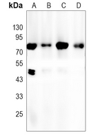 Western blot analysis of PKC beta expression in K562 (A), Jurkat (B), H1792 (C), mouse brain (D) whole cell lysates. (Predicted band size: 77 kD; Observed band size: 77 kD)
 -
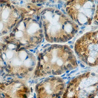 Immunohistochemical analysis of PKC beta staining in mouse colon formalin fixed paraffin embedded tissue section. The section was pre-treated using heat mediated antigen retrieval with sodium citrate buffer (pH 6.0). The section was then incubated with the antibody at room temperature and detected using an HRP conjugated compact polymer system. DAB was used as the chromogen. The section was then counterstained with haematoxylin and mounted with DPX.
 -
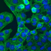 Immunofluorescent analysis of PKC beta staining in Hela cells. Formalin-fixed cells were permeabilized with 0.1% Triton X-100 in TBS for 5-10 minutes and blocked with 3% BSA-PBS for 30 minutes at room temperature. Cells were probed with the primary antibody in 3% BSA-PBS and incubated overnight at 4 °C in a hidified chamber. Cells were washed with PBST and incubated with a AF488-conjugated secondary antibody (green) in PBS at room temperature in the dark. DAPI was used to stain the cell nuclei (blue).