
 Datasheet
 MSDS
MSDSDescription:Rabbit polyclonal antibody to Caspase 3Immunogen:KLH-conjugated synthetic peptide encompassing a sequence within the center region of human Caspase 3. The exact sequence is proprietary.Purification:The antibody was purified by immunogen affinity chromatography.Clonality:PolyclonalForm:Liquid in 0.42% Potassium phosphate, 0.87% Sodium chloride, pH 7.3, 30% glycerol, and 0.01% sodium azide.Dilution:WB (1/500 - 1/1000), IH (1/100 - 1/200), IF/IC (1/100 - 1/500)Gene Symbol:CASP3Alternative Names:CPP32; Caspase-3; CASP-3; Apopain; Cysteine protease CPP32; CPP-32; Protein Yama; SREBP cleavage activity 1; SCA-1
Entrez Gene (Human):
836;
Entrez Gene (Mouse):
12367;
Entrez Gene (Rat):
25402;
SwissProt (Human):
P42574;
SwissProt (Mouse):
P70677;
SwissProt (Rat):
P55213;
Storage/Stability:Shipped at 4°C. Upon delivery aliquot and store at -20°C for one year. Avoid freeze/thaw cycles.
-
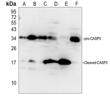 Western blot analysis of Caspase 3 expression in Hela (A), SGC7901 (B), EC9706 (C), mouse liver (D), mouse spleen (E), rat liver (F) whole cell lysates. (Predicted band size: 31 kD; Observed band size: 35; 17 kD)
 -
 Immunohistochemical analysis of Caspase 3 staining in human lung cancer formalin fixed paraffin embedded tissue section. The section was pre-treated using heat mediated antigen retrieval with sodium citrate buffer (pH 6.0). The section was then incubated with the antibody at room temperature and detected using an HRP conjugated compact polymer system. DAB was used as the chromogen. The section was then counterstained with haematoxylin and mounted with DPX.
 -
 Immunofluorescent analysis of Caspase 3 staining in HeLa cells. Formalin-fixed cells were permeabilized with 0.1% Triton X-100 in TBS for 5-10 minutes and blocked with 3% BSA-PBS for 30 minutes at room temperature. Cells were probed with the primary antibody in 3% BSA-PBS and incubated overnight at 4 °C in a humidified chamber. Cells were washed with PBST and incubated with a DyLight 594-conjugated secondary antibody (red) in PBS at room temperature in the dark. DAPI was used to stain the cell nuclei (blue).
 -
 Western blot analysis of Caspase 3 expression in HEK293T (A), HUT78 (B), MCF7 (C), mouse spleen (D), mouse brain (E), rat spleen (F), rat brain (G) whole cell lysates.
GRPR down-regulation inhibits spermatogenesis through Ca2+ mediated by PLCβ/IP3R signaling pathway in long-term formaldehyde-exposed rats
Therapeutic potential of mesenchymal stem cell-derived exosomal miR-296-5p and miR-337-3p in age-related erectile dysfunction via regulating PTEN
Piezo1 promotes intervertebral disc degeneration through the Ca2+/F-actin/Yap signaling axis