
 PIG3 Blocking Peptide
  Datasheet
 MSDS
MSDS
Description: The peptide is used to block Anti-PIG3 Antibody (#CPA5140) reactivity. Form: Lyophilized powder Gene Symbol: TP53I3 Alternative Names: PIG3; Quinone oxidoreductase PIG3; Tumor protein p53-inducible protein 3; p53-induced gene 3 proteinEntrez Gene (Human): 9540SwissProt (Human): Q53FA7Purity : >85%Directions for Use : Blocking Peptide to the diluted primary antibody in a molar ratio of 10:1 (peptide to antibody) and incubate the mixture at 4°C for overnight or at room temperature for 2 hours.Quality Control : The quality of the peptide was evaluated by reversed-phase HPLC and by mass spectrometry.Storage/Stability : Shipped at 4°C. Store at -20°C for one year.
-
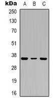 Western blot analysis of PIG3 expression in A549 (A), MCF7 (B), HepG2 (C) whole cell lysates.
 -
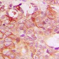 Immunohistochemical analysis of PIG3 staining in human breast cancer formalin fixed paraffin embedded tissue section. The section was pre-treated using heat mediated antigen retrieval with sodium citrate buffer (pH 6.0). The section was then incubated with the antibody at room temperature and detected using an HRP conjugacompact polymer system. DAB was used as the chromogen. The section was then counterstained with haematoxylin and mounted with DPX.