
 CBP20 Blocking Peptide
  Datasheet
 MSDS
MSDS
Description: The peptide is used to block Anti-CBP20 Antibody (#CPA4973) reactivity. Form: Lyophilized powder Gene Symbol: NCBP2 Alternative Names: CBP20; Nuclear cap-binding protein subunit 2; 20 kDa nuclear cap-binding protein; Cell proliferation-inducing gene 55 protein; NCBP 20 kDa subunit; CBP20; NCBP-interacting protein 1; NIP1Entrez Gene (Human): 22916Entrez Gene (Mouse) : 68092Entrez Gene (Rat) : 689116SwissProt (Human): P52298SwissProt (Mouse) : Q9CQ49SwissProt (Rat) : B1WC40Purity : >85%Directions for Use : Blocking Peptide to the diluted primary antibody in a molar ratio of 10:1 (peptide to antibody) and incubate the mixture at 4°C for overnight or at room temperature for 2 hours.Quality Control : The quality of the peptide was evaluated by reversed-phase HPLC and by mass spectrometry.Storage/Stability : Shipped at 4°C. Store at -20°C for one year.
-
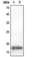 Western blot analysis of CBP20 expression in Hela (A), NIH3T3 (B) whole cell lysates.
 -
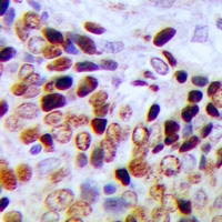 Immunohistochemical analysis of CBP20 staining in human prostate cancer formalin fixed paraffin embedded tissue section. The section was pre-treated using heat mediated antigen retrieval with sodium citrate buffer (pH 6.0). The section was then incubated with the antibody at room temperature and detected using an HRP conjugacompact polymer system. DAB was used as the chromogen. The section was then counterstained with haematoxylin and mounted with DPX.
 -
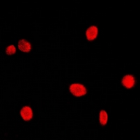 Immunofluorescent analysis of CBP20 staining in Hela cells. Formalin-fixed cells were permeabilized with 0.1% Triton X-100 in TBS for 5-10 minutes and blocked with 3% BSA-PBS for 30 minutes at room temperature. Cells were probed with the primary antibody in 3% BSA-PBS and incubated overnight at 4 °C in a hidified chamber. Cells were washed with PBST and incubated with a DyLight 594-conjugated secondary antibody (red) in PBS at room temperature in the dark. DAPI was used to stain the cell nuclei (blue).