
 Datasheet
 MSDS
MSDSDescription:Rabbit polyclonal antibody to pan methyl-lysineImmunogen:KLH-conjugated synthetic peptide of methyl-lysinePurification:The antibody was purified by immunogen affinity chromatography.Clonality:PolyclonalForm:Liquid in 0.42% Potassium phosphate, 0.87% Sodium chloride, pH 7.3, 30% glycerol, and 0.01% sodium azide.Dilution:WB (1/500 - 1/2000), IF/IC (1/50 - 1/200), IP (1/20 - 1/50)Alternative Names:Pan methyl-lysine
Storage/Stability:Shipped at 4°C. Upon delivery aliquot and store at -20°C for one year. Avoid freeze/thaw cycles.
-
 Western blot analysis of pan methyl-lysine expression in Hela (A) whole cell lysates. (Predicted band size: \; Observed band size: 15-60 kD)
 -
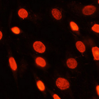 Immunofluorescent analysis of pan methyl-lysine staining in C6 cells. Formalin-fixed cells were permeabilized with 0.1% Triton X-100 in TBS for 5-10 minutes and blocked with 3% BSA-PBS for 30 minutes at room temperature. Cells were probed with the primary antibody in 3% BSA-PBS and incubated overnight at 4 °C in a humidified chamber. Cells were washed with PBST and incubated with a AF594-conjugated secondary antibody (red) in PBS at room temperature in the dark. DAPI was used to stain the cell nuclei (blue).