
 Datasheet
 MSDS
MSDSDescription:Rabbit polyclonal antibody to CD256Immunogen:KLH-conjugated synthetic peptide encompassing a sequence within the center region of human CD256. The exact sequence is proprietary.Purification:The antibody was purified by immunogen affinity chromatography.Clonality:PolyclonalForm:Liquid in 0.42% Potassium phosphate, 0.87% Sodium chloride, pH 7.3, 30% glycerol, and 0.01% sodium azide.Dilution:WB (1/500 - 1/1000), IH (1/100 - 1/200), IF/IC (1/100 - 1/500)Gene Symbol:TNFSF13Alternative Names:APRIL; TALL2; ZTNF2; Tumor necrosis factor ligand superfamily member 13; A proliferation-inducing ligand; APRIL; TNF- and APOL-related leukocyte expressed ligand 2; TALL-2; TNF-related death ligand 1; TRDL-1; CD256
Entrez Gene (Human):
8741;
Entrez Gene (Mouse):
69583;
SwissProt (Human):
O75888;
SwissProt (Mouse):
Q9D777;
Storage/Stability:Shipped at 4°C. Upon delivery aliquot and store at -20°C for one year. Avoid freeze/thaw cycles.
-
 Western blot analysis of CD256 expression in HEK293T (A), Hela (B), H446 (C), mouse brain (D), mouse kidney (E), rat brain (F), rat kidney (G) whole cell lysates. (Predicted band size: 22; 24; 25; 27; 36 kD; Observed band size: 22 kD)
 -
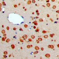 Immunohistochemical analysis of CD256 staining in human brain formalin fixed paraffin embedded tissue section. The section was pre-treated using heat mediated antigen retrieval with sodium citrate buffer (pH 6.0). The section was then incubated with the antibody at room temperature and detected using an HRP conjugated compact polymer system. DAB was used as the chromogen. The section was then counterstained with haematoxylin and mounted with DPX.
 -
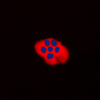 Immunofluorescent analysis of CD256 staining in H9C2 cells. Formalin-fixed cells were permeabilized with 0.1% Triton X-100 in TBS for 5-10 minutes and blocked with 3% BSA-PBS for 30 minutes at room temperature. Cells were probed with the primary antibody in 3% BSA-PBS and incubated overnight at 4 °C in a humidified chamber. Cells were washed with PBST and incubated with a DyLight 594-conjugated secondary antibody (red) in PBS at room temperature in the dark. DAPI was used to stain the cell nuclei (blue).
Factors supporting intrathecal humoral responses following viral encephalomyelitis
High expression of TNFSF13 in tumor cells and fibroblasts is associated with poor prognosis in non-small cell lung cancer
Lactobacillus rhamnosus GG Promotes Early B Lineage Development and IgA Production in the Lamina Propria in Piglets