
 Datasheet
 MSDS
MSDSDescription:Recombinant rabbit monoclonal antibody to PDC-E2Immunogen:KLH-conjugated synthetic peptide encompassing a sequence within human PDC-E2 protein. The exact sequence is proprietary.Purification:The antibody was purified by immunogen affinity chromatography.Clonality:MonoclonalForm:Liquid in PBS, pH 7.4, containing 50% glycerol, 0.2% BSA and 0.01% sodium azide.Dilution:WB (1/500 - 1/1000), IF/IC (1/50 - 1/200)Gene Symbol:DLATAlternative Names:DLTA; Dihydrolipoyllysine-residue acetyltransferase component of pyruvate dehydrogenase complex, mitochondrial; 70 kDa mitochondrial autoantigen of primary biliary cirrhosis; PBC; Dihydrolipoamide acetyltransferase component of pyruvate dehydrogenase complex; M2 antigen complex 70 kDa subunit; Pyruvate dehydrogenase complex component E2; PDC-E2; PDCE2
Entrez Gene (Human):
1737;
Entrez Gene (Mouse):
235339;
Entrez Gene (Rat):
81654;
SwissProt (Human):
P10515;
SwissProt (Mouse):
Q8BMF4;
SwissProt (Rat):
P08461;
Storage/Stability:Shipped at 4°C. Upon delivery aliquot and store at -20°C for one year. Avoid freeze/thaw cycles.
-
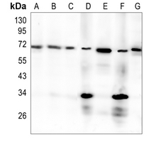 Western blot analysis of PDC-E2 expression in K562 (A), PC3 (B), HepG2 (C), mouse kidney (D), mouse muscle (E), rat kidney (F), rat muscle (G) whole cell lysates. (Predicted band size: 68 kD; Observed band size: 69 kD)
 -
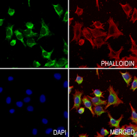 Immunofluorescent analysis of PDC-E2 staining in A375 cells. Formalin-fixed cells were permeabilized with 0.1% Triton X-100 in TBS for 5-10 minutes and blocked with 3% BSA-PBS for 30 minutes at room temperature. Cells were probed with the primary antibody in 3% BSA-PBS and incubated overnight at 4 °C in a hidified chamber. Cells were washed with PBST and incubated with a AF488-conjugated secondary antibody (green) in PBS at room temperature in the dark. Phalloidin - AF594 was used to stain Actin filaments (red). DAPI was used to stain the cell nuclei (blue).
Orchestrated Cu2+-coordinated tetracycline-porphyrin self-assembly remodels tumor microenvironment for photo-enhanced immuno-chemodynamic therapy