
 Datasheet
 MSDS
MSDSDescription:Recombinant rabbit monoclonal antibody to NDUFS2Immunogen:KLH-conjugated synthetic peptide encompassing a sequence within human NDUFS2 protein. The exact sequence is proprietary.Purification:The antibody was purified by immunogen affinity chromatography.Clonality:MonoclonalForm:Liquid in PBS, pH 7.4, containing 50% glycerol, 0.2% BSA and 0.01% sodium azide.Dilution:WB (1/500 - 1/1000), IF/IC (1/50 - 1/200)Gene Symbol:NDUFS2Alternative Names:NADH dehydrogenase [ubiquinone] iron-sulfur protein 2, mitochondrial; Complex I-49kD; CI-49kD; NADH-ubiquinone oxidoreductase 49 kDa subunit
Entrez Gene (Human):
4720;
Entrez Gene (Mouse):
226646;
Entrez Gene (Rat):
289218;
SwissProt (Human):
O75306;
SwissProt (Mouse):
Q91WD5;
SwissProt (Rat):
Q641Y2;
Storage/Stability:Shipped at 4°C. Upon delivery aliquot and store at -20°C for one year. Avoid freeze/thaw cycles.
-
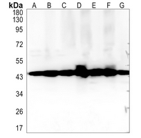 Western blot analysis of NDUFS2 expression in MCF7 (A), Jurkat (B), K562 (C), mouse kidney (D), mouse muscle (E), rat kidney (F), rat muscle (G) whole cell lysates. (Predicted band size: 52 kD; Observed band size: 45 kD)
 -
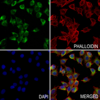 Immunofluorescent analysis of NDUFS2 staining in A375 cells. Formalin-fixed cells were permeabilized with 0.1% Triton X-100 in TBS for 5-10 minutes and blocked with 3% BSA-PBS for 30 minutes at room temperature. Cells were probed with the primary antibody in 3% BSA-PBS and incubated overnight at 4 °C in a hidified chamber. Cells were washed with PBST and incubated with a AF488-conjugated secondary antibody (green) in PBS at room temperature in the dark. Phalloidin - AF594 was used to stain Actin filaments (red). DAPI was used to stain the cell nuclei (blue).