
 Datasheet
 MSDS
MSDSDescription:Recombinant rabbit monoclonal antibody to EGLN2Immunogen:KLH-conjugated synthetic peptide encompassing a sequence within human EGLN2 protein. The exact sequence is proprietary.Purification:The antibody was purified by immunogen affinity chromatography.Clonality:MonoclonalForm:Liquid in PBS, pH 7.4, containing 50% glycerol, 0.2% BSA and 0.01% sodium azide.Dilution:WB (1/500 - 1/1000), IF/IC (1/50 - 1/200)Gene Symbol:EGLN2Alternative Names:EIT6; Egl nine homolog 2; Estrogen-induced tag 6; HPH-3; Hypoxia-inducible factor prolyl hydroxylase 1; HIF-PH1; HIF-prolyl hydroxylase 1; HPH-1; Prolyl hydroxylase domain-containing protein 1; PHD1
Entrez Gene (Human):
112398;
Entrez Gene (Mouse):
112406;
SwissProt (Human):
Q96KS0;
SwissProt (Mouse):
Q91YE2;
Storage/Stability:Shipped at 4°C. Upon delivery aliquot and store at -20°C for one year. Avoid freeze/thaw cycles.
-
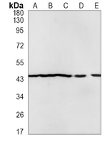 Western blot analysis of EGLN2 expression in Hela (A), K562 (B), THP1 (C), mouse liver (D), mouse muscle (E) whole cell lysates. (Predicted band size: 43 kD; Observed band size: 44 kD)
 -
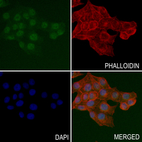 Immunofluorescent analysis of EGLN2 staining in MCF7 cells. Formalin-fixed cells were permeabilized with 0.1% Triton X-100 in TBS for 5-10 minutes and blocked with 3% BSA-PBS for 30 minutes at room temperature. Cells were probed with the primary antibody in 3% BSA-PBS and incubated overnight at 4 °C in a hidified chamber. Cells were washed with PBST and incubated with a AF488-conjugated secondary antibody (green) in PBS at room temperature in the dark. Phalloidin - AF594 was used to stain Actin filaments (red). DAPI was used to stain the cell nuclei (blue).